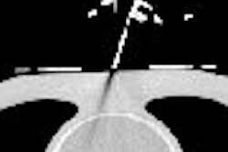
VIENNA - When it comes to lung biopsies, the key factors are patient consent and collaboration, the indications and contraindications, imaging review, guidance selection, patient positioning and the use of local anesthesia, and recovery and potential complications, a leading Italian interventional radiologist told a standing-room only Mini Course on Thursday.
"Talking to the patient is the single most important thing when you do a lung biopsy," said Dr. Mario Bezzi, from the department of radiological sciences at the Sapienza University of Rome. "Remember to team up with the patient."
 Dr. Mario Bezzi from Rome.
Shallow breathing for upper lesions is important, and having the patient in a prone position reduces lung excursion. Clear advice on following instructions is vital, even if pressure or pain is felt, he added.
Bezzi had some practical advice on injection of contrast during the procedure to differentiate the lesion from the vascular structure.
"I start the procedure without contrast, based on the previous imaging. I can place the contrast on the noncontrast images, but when it comes to going near the vessel, I inject contrast medium at that point. I split the dose of the contrast medium if I need more bolus during the procedure," he said. "For a skinny lady, I would say that boluses of around 30 mL of contrast media will show you the vessels. For larger gentlemen, I would use 40 to 45 mL. At that point, you can be sure that you have three to four boluses."
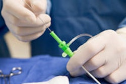
A point of controversy, however, was needle size. A member of the audience said he uses a 20G Tru-Cut side-cutting needle for most lung biopsies and gets good results, but Bezzi expressed surprise. He prefers 20G modified Menghini end-cutting needles because compared with side-cutting needles, modified Menghini devices allow for a bigger tissue core for each sample.
Use the coaxial technique to obtain multiple samples with a single pleural puncture; an introducer needle carries a smaller needle, and increases needle size by 1G (e.g., 17G carries 18G), he commented.
Immediate postprocedure care should include bed rest; supine or lateral decubitus (45°); O2, pulse, and blood pressure monitoring; between one and four hours for the patient in a dedicated recovery room; and a chest x-ray after four hours or if there is any clinical deterioration. Many interventional radiology services perform lung biopsy as a day case, with discharge after four hours, and readmission only if certain symptoms persist, according to Bezzi.
A pneumothorax is bound to happen at some point and is something everyone must face up to, he said. The main risk factors are chronic obstructive pulmonary disease, patient age, needle size, lesion size less than 10 mm, lesion depth, lack of previous surgery, procedure time, an operator's experience, and conditions that increase chest pressure, such as a cough or tracheal cannula.
Preprocedural evaluation for lung parenchyma comprises peripheral emphysematous bullae, bronchiectasis, and the presence of comorbidities and conditions that can suddenly increase pressure in the chest.
Other potential complications are hemoptysis, hemorrhage, failure and the consequent need to repeat the procedure, infection/sepsis, and air embolism, he said. A hemorrhage is very often asymptomatic, self-limiting, and intraparenchymal. In the case of hemoptysis, it's necessary to reassure the patient and inform a referring physician of the need for closer monitoring during the recovery period.
An air embolism can occur if a vessel is punctured inadvertently, and air is aspirated by negative pressure or embolized by positive pressure. The symptoms resemble those of stroke, transient ischemic attack, seizures, or cardiopulmonary collapse, and a CT scan of the chest and brain is recommended. To manage air embolism, make the diagnosis and place the patient in the left lateral decubitus position or with the head down, and 100% O2 or hyperbaric O2 should be administered in the intensive care unit, he noted.
"You held this excellent lecture in English, but you used gauge numbers. At home in Italy, do you use metric units?" asked a delegate.
"We use gauge because otherwise we would not understand each other," Bezzi replied. "G is the standard name. Give me a 20G and everybody understands what you want."
Originally published in ECR Today on 7 March 2014.
Copyright © 2014 European Society of Radiology


