VIENNA - The ultrademanding and highly competitive nature of modern professional sport was visible to all during Sunday's refresher course on overuse injuries.
Stress reactions are the most important lesions to recognize in the gymnast's spine, and although there is no consensus about imaging, plain films and MRI, plus CT in selected cases, are the best option, said Dr. Milko De Jonge, from the department of radiology at Zuwe Hofpoort Hospital in Woerden, the Netherlands.
 Dr. Milko De Jonge from Woerden, the Netherlands. Image courtesy of the European Society of Radiology.
The term "gymnastics" derives from the Greek word "γυμνóς," and when the Ancient Greeks engaged in the activity, they were naked. "I wonder how many gymnasts today would take part in it if they had to be naked. Probably not very many of them -- I know I wouldn't!" he joked.
From a medical perspective, a major problem is that gymnasts are so young when they start participating. In the Dutch competition league, gymnasts are as young as 6 years old, and they are considered to be veterans at 18. There are 245,000 members of the country's gymnastics union, which represents a slight decline since 2010, stated De Jonge. As many as 20 million women in the U.S. take part in gymnastics, so participation remains healthy in most parts of the world. It became an Olympic sport for men in 1896 and for women in 1928.
"Low back pain in gymnasts is common. It originates predominantly from hyperextension and rotation, with axial forces," he said. "Disk disease is also common."
Not all disk disease is pathognomonic for Scheuermann's disease, however. To avoid causing unnecessary anxiety to patients, he recommends avoiding the term Scheuermann's disease, except for classical cases, and there is no room for three-quarter views.
The frequency and intensity of training are high. Up to the age of 14, talented youngsters train for between 15 and 25 hours a week, while national team members train for around 35 hours a week, De Jonge said.
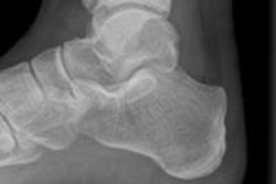
In professional football players, overuse injuries in the ankle are particularly common, according to Dr. Patricia Cunningham, a radiologist from Louth-Meath Hospital Group in Ireland. She said it's important to diagnose injuries of the ankle ligaments, including sequelae such as anterolateral impingement, as well as injuries of tendons around the ankle and bone and cartilage injuries in the ankle, including anterior and posterior impingement.
Golfers tend to suffer most often from upper limb ailments, and radiologists need to have an understanding of the swing mechanics to appreciate the asymmetrical nature of golf injury, according to Dr. Philip O'Connor, a musculoskeletal radiologist from Leeds Teaching Hospitals and the University of Leeds, U.K.
In a recent survey of players on the European Tour, the results of which are due to be published soon by the British Journal of Sports Medicine, the wrist accounted for 35% of total injuries, of which 30% were previous injuries. A total of 128 questionnaires were completed, representing a response rate of 84%. The golfer's lead wrist was injured in 67% of cases, and of those injuries, 35% were ulnar-sided, 11% were radial-sided, 33% were dorsal, and 21% were other.
Fractures of the hook of the hamate, also called the hamulus, are the most common fracture in a golfer, and are usually stress fractures, said O'Connor, who led the imaging team at the 2012 Olympics in London. The nondominant wrist can be affected, and they are difficult to diagnose clinically and are occult on conventional wrist radiographs.
In sports injuries encountered at Leeds, radiologists spend a lot of time trying to differentiate between established defects and new acute stress fractures, he said. For instance, a 24-year-old cricketer might have developed his defects at the age of 14 of 15, he commented.
Originally published in ECR Today on 11 March 2013.
Copyright © 2013 European Society of Radiology


