VIENNA - Developers of MRI systems are working hard to find new features that will convince customers to look twice at their products when considering investing in their technologies. A wide variety of developments designed to improve productivity and enhance patient care is on show at this year's European Congress of Radiology (ECR) technical exhibition.
Siemens believes that time and effort is often wasted when carrying out advanced imaging procedures in looking at parts of the body that are of little or no interest to the referring physician as they are irrelevant to the condition under investigation. That is because a traditional MRI unit will collect data in a slice through the whole human body when the reason for the examination is to look at a single organ or tissue.
The company's solution to this problem is an innovation that is being shown for the first time in Europe here in Vienna. It aims to make it available for its top-of-the-range Magnetom Skyra 3T system before the end of the year.
The TimTX TrueShape is a system architecture for parallel transmit (pTX) technology that is being introduced to complement its TimTX TrueForm magnet and gradient design, according to Christof Zindel, head or product definition and marketing for Siemens' MRI business. TimTX TrueForm made its debut at ECR in 2007 as a system designed to optimize the signals received and minimize distortion at the edge of the field-of-view. TimTX TrueShape focuses on an earlier stage of the process by controlling the nature of the signals produced. Instead of a single radiofrequency pulse at a fixed amplitude, as in conventional MRI, the system produces multiple signals shaped to the specific needs of a particular clinical application.
 |
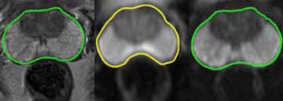
| Early results from a prostate cancer study using Syngo ZOOMit. The spatial resolution was increased in the phase-encoding direction, leading to 1.6-mm squared in-plane resolution at 3-mm slice thickness in imaging the highly specific diffusion microstructure. The case demonstrates another property of zoomed echo-planar imaging (EPI): minimization of image distortions. The same green contour is laid over the anatomical image and the zoomed EPI image, showing a good match. Once the contour is extracted on a conventional image, there are clear differences due to the air/tissue interfaces and the resulting susceptibility artifacts. (Provided by Siemens) |
At ECR, Siemens is unveiling the first application of this parallel transmit technology, which it is calling syngo ZOOMit. It is capable of selective excitation to allow the radiographer to shape the image produced and focus attention on a particular region of the body, organ, or even a single anatomical feature.
"It is all about selective excitation. We can create the signal to produce excitation in the protons within a specific tissue, such as the prostate or heart, without involving the surrounding tissue. That is important because the surrounding tissues can often be the source of infolding and other artifacts in the image that is produced," Zindel explained. "By limiting the excitation lines and the read out to the specific organ rather than the whole field-of-view, you can reduce the scan time by up to 45%, which will further improve the image quality by minimizing motion artifacts."
Cutting down on unnecessary data acquisition and processing from the adjacent organs has also been shown to produce diagnostically significant improvements in image quality and increased spatial resolution. He adds that the shorter examination time will provide benefits in workflow for the radiology department and reduce the stress experienced by its patients. He also highlights the versatile nature of the parallel transmission technology that forms the basis for the new application.
 |
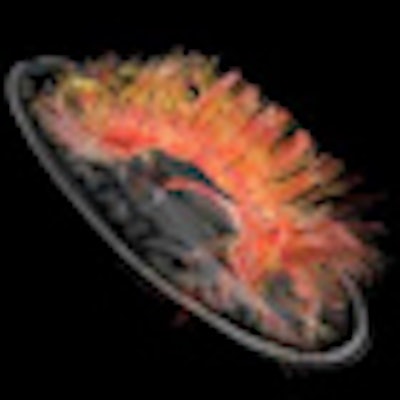
| Diffusion tensor fiber-tracking brain image overlaid on Bravo and color orientation, using a Discovery MR750w 3-tesla system. (Provided by GE) |
"Tim TX is a program, not a one-off project. So syngo ZOOMit is the first product of the TrueShape technology," Zindel noted. "We have a clear route open to develop new applications and are confident that these will be available to be shown at future events."
Hitachi is presenting Echelon Oval, which has an oval bore. The company reasons that the human body is not circular in cross section, so why should the scanner be? It has redesigned the system to be wider in the horizontal than the vertical plane, with a 74-cm-wide bore that reportedly provides more room than any other 1.5-tesla machine to accommodate even most broad-shouldered patients. This feature will be appreciated by athletes or bariatric patients, as well as anxious individuals who may experience claustrophobic feelings within a standard-sized bore. Furthermore, this shape also facilitates off-centre imaging of more peripheral body areas such as the shoulder.
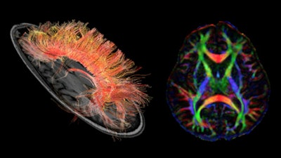
 Diffusion tensor imaging (DTI) tractography evaluation in the C-spine. Syngo DTI tractography uses diffusion tensor data and allows 3D visualization of specific white-matter tracts, e.g., to determine the location of the corticospinal tract or the thalamocortical tract. (Provided by Siemens)
In designing the new system, Hitachi has also aimed to improve the technologist's experience. The company's recently introduced Workflow Integrated Technology (WIT) system helps technologists overcome the inconvenience of removing and selecting coils so that more of their time is spent on scanning and looking after their patients. Another advantage is magnet monitoring. The technologist can input patient data and check ECG and respiratory sensing without having to move across to the control workstation, which reduces procedure time and has a positive impact on workflow. The new unit also has a number of other advanced features, such as the AutoPose Brain application for automatic slice selection, which reduces the number of steps needed by the technologist and increases the consistency of slice placement.
Improved patient comfort was a key priority for GE Healthcare in designing the latest version of its flagship 3-tesla MRI unit, the Discovery MR750w. The 70-cm-bore unit features a compact superconducting magnet and delivers a full 50 x 50 x 50-cm field-of-view (FOV), well-suited for large FOV imaging and off-centre imaging. It also features the Geometry Embracing Method (GEM) suite of lightweight flexible coils with anatomy-optimized element geometry, so that it can accommodate patients of different size and shape, and providing excellent RF and signal homogeneity to make the best use of the available FOV, according to GE.
The GEM system is designed to reduce coil handling and simplify patient setup, and to adapt to the patient's contours so that the coil elements are optimally positioned relative to the shape of the individual. The system has 160 coil mode configurations and can automatically select the coil elements best suited to image the prescribed FOV for that particular clinical indication. GE estimates that the GEM suite can be deployed in up to 98% of exam types. Other features include a total 2,056-cm scanning range, feet-first scanning for nearly all exams, and improved padding on the patient table to help minimize pressure points and discomfort.
 |
| Left, BSI (blood sensitive imaging) is used to acquire high-contrast and high-resolution images in a short time based on different T2-weighted contrast of tissues. (Provided by Hitachi) Right, CSI (chemical shift imaging) case of pediatric glioma seen at Iwate Medical University, Japan. (Provided by Hitachi) |
A highlight at last year's ECR exhibition were the Philips Ingenia 1.5- and 3-tesla MRI systems, described as the first ever digital broadband MRI technology. The signal is digitized in the coil immediately adjacent to the patient, delivering gains in both signal-to-noise ratio (SNR) and imaging acceleration. The company returns this year with various updates to this core technology, developed in collaboration with some of the growing band of hospital centers that have installed the system.
"Ingenia delivers up to 40% more SNR, and has the largest field-of-view in the industry, and this is relevant in the light of the diversification of MRI from the traditional applications in neurology towards whole-body oncology and cardiology scans," said Jurnjan van den Bremer, international business manager for Philips' MR activities. "Ingenia accelerates patient management, through advanced coil solutions and smart assist features, with 30% increase in throughput. Ingenia includes a flex coverage anterior coil, hidden in the table, requiring virtually no coil handling in about 60% of cases."
The new features on display at ECR 2012 include an application that enables the time needed for whole-body imaging studies to be reduced from around one hour down to 10 minutes using the coronal diffusion-weighted whole-body imaging with background body signal suppression technology. The latest Philips technology also includes the IntelliSpace portal, allowing the user to gain access to images anywhere for advanced visualization of the data.
Originally published in ECR Today March 3, 2012.
Copyright © 2012 European Society of Radiology


