The current generation of high-field MR systems (7-tesla and higher) is not ready for clinical use because radiofrequency coils are not available, protocols have not been optimized, and current systems are expensive and need a lot of space. However, future 7-tesla magnets look set to be partially shielded, and will require significantly less space and shielding measures. Emerging technologies such as parallel radiofrequency transmission will address another important problem: inhomogeneities in the transmit field, which cause susceptibility artifacts.
These are the opinions and predictions of Dr. Michael Bock, a medical physicist at the German Cancer Research Center (Deutsches Krebsforschungszentrum, DKFZ) in Heidelberg, which is the largest biomedical research institute in Germany.
"High-resolution MRI in the brain, and in other anatomical areas (e.g., the knee), will become the first established clinical applications of high-field MRI," he said. "For example, we are able to visualize the tiny neovasculature in brain tumors, so high-field MRI might be a powerful tool to assess therapy response to the more and more popular neoangiogenic treatment," he commented.
The use of nuclei other than protons (sodium-23, oxygen-17, phosphorus-31) will open up new possibilities for assessing metabolic information, but there are still many technical problems to be solved, particularly in relation to the high and localized energy deposition, he warned. The higher energy deposition is hard to predict because local focusing of the radiofrequency fields can occur.
The trend to ever higher fields in MRI is typically motivated by the gain in spatial resolution that can be achieved with high-field MR systems, but with increasing field strength, several problems arise, including new image artifacts. High-field MRI will not provide a meaningful solution for each and every imaging problem, and the general radiologist should be aware of this when deciding which field strength to choose, Bock stated.
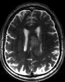 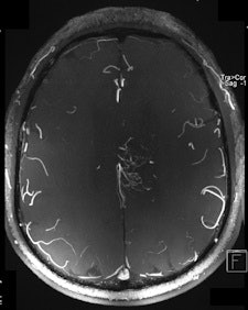 |
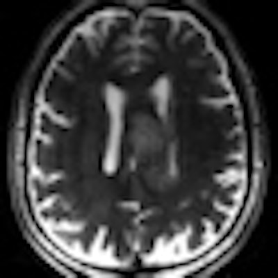
| Two 7-tesla images of a glioblastoma patient. Left: T2-weighted image shows brain lesion with very high spatial resolution. The heterogeneity in the lesion is already visible. Right: Time-of-flight MR angiogram shows the arterial vessels at the periphery of the lesion. The irregular vessel structure indicates neoangiogenesis, and it would be very interesting to see whether this vascular structure changes under new forms of neoangiogenic therapy. Image courtesy of Dr. Michael Bock. |
Noise is another issue. The forces acting on the imaging gradients increase linearly with field strength, and so an increase in sound pressure occurs at higher fields due to the stronger vibration of the gradient coils. Aware of this challenge, MR manufacturers have included additional sound protection measures, such as more rigid gradient coils or better damping, into their systems. In the 7-tesla unit at the DKFZ, sound pressure levels are now similar to those at 3-tesla. Nevertheless, passive sound protection using earplugs or muffs is always indicated at high-field strengths, he said.
The need for sound protection measures and the requirement for stronger gradient systems do not appear to have a substantial impact on overall costs. The major cost comes from the magnet, and the gradient redesign does not add massively to the total system cost, Bock maintained.
When the field strength rises, the signal-to-noise ratio (SNR) also increases linearly. Since MRI is intrinsically an insensitive technology, every increase in SNR is more than welcome. At a higher SNR, it is possible to lower the voxel size (i.e., increase the spatial resolution), acquire images faster, and detect MR signals from rare nuclei such as sodium, he pointed out.
Possible clinical applications of 7-tesla include the detection of very small lesions such as metastases, near-real-time imaging of the heart, and imaging of cell vitality using sodium MRI. Furthermore, Bock notes that the increased susceptibility contrast is widely used for neurofunctional MRI (fMRI).
Operating at increased magnetic fields makes it easier to obtain T2* contrast-enhanced images and improved implementation of susceptibility-weighted imaging, in which the phase of gradient-echo images provides information about local variation of magnetic susceptibility, according to Richard Bowtell, a professor from the Magnetic Resonance Centre, School of Physics and Astronomy, at the University of Nottingham in the U.K. In the brain, such variation appears to be dominated by differences in iron concentration and myelin content, so that high-field SWI may provide useful information about the progression of neurodegenerative disease. The elevated T1 relaxation times at 7-tesla also offer benefits for arterial spin labeling and time-of-flight angiography.
In his ECR presentation, Bowtell discussed the current and potential future applications of high-field MRI in clinical and preclinical studies in a number of areas, along with the barriers to wider usage of 7-tesla systems for clinical studies.
Originally published in ECR Today March 6, 2011.
Copyright © 2011 European Society of Radiology


