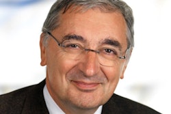
Presiding over this year's European Congress of Radiology (ECR) will be Yves Menu, MD, the French gastrointestinal specialist. For his day job, Menu faces the equally daunting task of reorganizing the department of radiology at Saint Antoine Hospital in Paris.
Shortly after his arrival at Saint Antoine in 2008, Menu was handed what he calls a Rubik's Cube of issues to solve. His radiology group consolidated with three other hospitals that now form the East University Hospital Group within the massive organization, Assistance Publique-Hôpitaux de Paris (AP-HP).
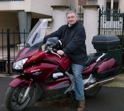 Revving up for action in Vienna: Motorbiking is Prof. Menu's newest passion, and he learned to ride last year.
Also a professor of radiology at the University of Paris, Menu brings extensive experience to this new puzzle. During the past 20 years he led radiology services at prestigious institutions that include Bichat Hospital, Beaujon Hospital, and Bicêtre Hospital.
AuntMinnieEurope.com caught up with Menu for a conversation on the eve of this year's ECR.
AuntMinnieEurope: How will Saint Antoine emerge from its consolidation with the Rothschild, Tenon, and Trousseau hospitals?
Yves Menu: To this point, Saint Antoine and the other three hospitals have been defined by their walls. The head of each service has been his own boss and in many areas we have been competing. This consolidation creates a complementary and not competitive care model.
Trousseau is a pediatric hospital and Rothschild is a rehabilitation care center, both of which are supported by radiology services with imaging resources specifically adapted to their patients. Today we tend to compete in pneumonology, women's health, neurology, cardiology, and emergency. Under the new organization, Saint Antoine will be the specialized center for cardiology and neurology, while Tenon will offer pneumonology, gynecology, and maternity.
Everyone understands the necessity for this change. The difficulty lies in the logistics, which create a Rubik's Cube during the transition period that will be tricky to direct. Working together does not necessarily mean mixing these specialties -- the interface between these groups is always variable.
Will the change bring new capabilities to Saint Antoine?
Currently we have two 64-slice CT scanners, two 1.5-tesla MRI systems, three ultrasound scanners, and several rooms for conventional radiography. We also have an interventional angiography room. With the cardiology focus, we will build a second cath lab.
I see the demand for our CT scanners diminishing. We added the second scanner a year ago because demand was surpassing our capacity. Yet already we find the workflow is quite well-aligned with demand. The waiting time for a CT is one hour here. CTs are now so rapid they easily handle 50 to 60 exams per day, even as I see a reduced requirement for these exams.
On the other hand, the waiting time for MRIs is increasing. Right now both MRI units run full time, so expanding the neurology practice is going to challenge that capacity -- but I expect to add a 3-tesla MRI system.
There is an acute shortage of scanners in France, which results in long waiting lists for exams. Is this true for your operation?
The situation in France is not good and it is particularly troubling for MRI exams. The shortage of MRIs varies greatly across the country, with the situation a bit better in the Paris region.
As the head of radiology for Saint Antoine, my concerns are maintaining our uptime and filling available exams slots. I have a capacity to conduct 50 MRI exams per day. The demand is for 80 exams per day.
Our waiting times are actually very short because we manage the upstream demand by working with referring physicians to ensure their requests are justified and that they will add value. It is the best way to ensure the quality of our service. We want patients to come in while they still have the condition that needs to be examined, not 30 days later.
We also call patients the day before to ensure they are coming. It takes effort and time but it is worth it because otherwise we have two or three people each day who fail to show up for their exam. If we have 24 hours' notice, we always can find someone to come in and fill the slot.
Where the demand remains greater than our capacity, patients are referred to other centers. Here I have concerns that are national rather than local. I cannot be satisfied with the current choices, which are either to send patients to other centers, or else stretch out the waiting times for exams. Also most centers -- under the pressure of an increasing waiting time -- are forced to work fast and not necessarily at the level of quality they are normally capable of delivering. The question of quality, or 'scientific added value' if you like, should be prominent, rather than the calibration of resources only to the quantity. Pediatrics is an excellent example. It takes time to perform an MRI exam in children for many reasons that a radiologist understands. A radiologist, yes, but not necessarily an administrator, who looks only at the numbers and reimbursement.
It has been reported that only 20% of French public hospitals are equipped with PACS. What is the situation at Saint Antoine?
We have a good PACS here and it is connected to an AP-HP network for all of Paris. Five years ago we had nothing and now we have such a good system that I have trouble remembering how we worked without it.
The disadvantage is readings become highly individualized and it is more difficult to share a reading. It is like someone zapping channels on the television with the remote control. The navigation becomes highly individualized and the person next to the remote, without the same visual rhythm, is frustrated.
The number of images has grown incredibly, to thousands of images for an exam. Even the idea of an "image" no longer exists because it does not mean anything by itself. Each image is nothing more than a letter in an alphabet. These letters form words then sentences but they no longer have a value alone.
The radiology report with a fixed object for viewing also no longer makes sense. A hypertext link to a series of images tagging segments is what makes sense. Instead of reporting a tumor in the right segment, we send the segment in a volume, a multimedia document, not an image. Once linked with this selected region of interest, the reader can move through the slices.
Software is bringing tremendous changes to radiology . . .
Yes, but medicine has changed as well, requiring things traditionally not asked of us. Twenty years ago, hardly 20% of patients required a follow-up exam. Nor was there a need to review the same patient across different exams on multiple modalities.
Today 80% of the patients in a university hospital setting come back for a second session, so the comparison of images from previous sessions has become extremely significant. The reason is simple: the advances in oncology with expanding types of chemotherapy treatments and increasing patient survival rates.
Twenty years ago, a patient diagnosed with colon cancer survived seven months on average. Now it is 32 months. If we are asked to perform a scan every three months, you can see how the history grows. Twenty years ago there was one chemotherapy drug. If it failed, there was not any interest in performing subsequent scans. Today when the first treatment fails, there is a second therapy, then a third.
Each time there is a need for medical imaging -- every three months. The number of patients with colon cancer has not changed appreciably in 20 years. But the number of scans per patient with colon cancer has multiplied by 10.
Doesn't computer-aided detection (CAD) help?
CAD is a welcome improvement, even essential. We have had such tools for a bit more than two years and we use them every day. There is a high sensitivity but a low specificity. It is not revolutionary but it is certainly a time-saver.
CAD raises a larger issue, which is delegating work. We need to distinguish where the experienced radiologist adds a real value and where, with new tools, perhaps we do not add anything especially valuable, such as reading follow-up exams for cancer patients. If a trained radiographer with CAD is capable of doing something as well as a radiologist, then this task should be delegated to the radiographer.
In detection for follow-up, perhaps it is not necessary to know the mechanism of action between the cells of a neoplasia just to be able to spot a nodule in the colon. On the other hand, in verifying the detected nodules, to identify associated structures or to know what strategy the detection of this nodule implies, then yes, medical studies are critical. I prefer radiologists apply themselves for five minutes to verify the results of the follow-up exam rather than spending the time it takes to look through the entire exam.
Doesn't this pose a threat to the traditional role of the radiologist?
Radiologists need to become true clinicians and we should not fear the idea of clinicians becoming radiologists. More and more the separation between these practices is becoming a porous membrane. Medical practice needs to move closer to the models we see today with breast cancer, which places in the same center a radiologist, an oncologist, a surgeon, a radiotherapist, and an anatomical pathologist. The patient comes in with a problem and leaves the same day with a solution.
For this to be effective, the clinician needs to know more about radiology, and the radiologist needs to understand more about the clinical issues. Medicine can no longer be each of us in our little corner doing a good job but isolated from the rest of the process. We cannot allow situations where an image is lost for three months in the files or patients are not followed or do not return for appointments. We need to completely reorganize our way of working.
At ECR 2011 you will moderate a session on virtual colonoscopy versus optical colonoscopy. What can we expect to hear from you as a gastrointestinal specialist on this topic?
We all know that scientifically the two methods are far more complementary than competitive. There is reimbursement to consider and commercial issues can come into play, but virtual colonoscopy is a very good method. Clearly it does not have the advantages of an optical examination for sensitivity and specificity.
In setting policy there is no question we can utilize virtual colonoscopy for screening but only optical colonoscopy is effective for characterizing lesions. At the same time, when there is a patient with a low probability of having cancer, using an optical colonoscopy is like taking a hammer to swat a fly. Patients don't like it, clinicians don't like it. No one likes it.
Gastroenterology is a special focus for ECR 2011. How do you view the relationship between radiology and your specialty in gastroenterology?
When you look at the history of the links between radiology and gastroenterology, it is exemplary. Classic digestive radiology was destroyed by endoscopy. But overall, there has been very good communication and ongoing exchange between the two specialties as we have respected the progress. There are debates, sometimes quite heated, but overall we have the same objectives medically and scientifically.
It is like a very good marriage with occasional disputes, but gastroenterology has proved to be a unifying specialty while radiology is a practice that is open to sharing. So it has been a natural, easy marriage.
The strongest proof is when the European gastronomical societies restructured to federate as the United European Gastroenterology Federation; the European Society of Gastrointestinal and Abdominal Radiology (ESGAR) joined this federation as a member.
What lessons do you take away from your year as president of ECR 2011?
The question I ask is what is the role of these congresses in a world where each one in his service has so much work and there are so many new means for sharing information?
I wonder if we no longer had the support of industry through sponsoring, would these congresses continue. These medical congresses are so heavily sponsored directly but also indirectly with companies supporting research and investigators. I wonder about all the jet fuel that is spent to bring us to these congresses.
In the future why should we continue? If congresses do not change their format and their reason for being, will there continue to be an interest in coming? Why can we not participate over the Internet with these presentations? So many of us already do.
I have people who write to me saying they reviewed my recent presentation on a Sunday evening and they have some questions. Each week, when I have a spare moment, I find myself reviewing presentations on the Internet. It is learning when you want, where you want. Sometimes I almost fall asleep reading some of them but also there are moments where I discover unexpected things.
Moving forward, medical congresses need to add something different, bring something new. In two words, that needs to be "interactivity," and secondly, "multidisciplinary."
Interactivity means that congresses bring together people physically for a direct dialogue and not a prepared dialogue, a discussion that is over organized. For this reason at this year's ECR, we have included an enormous number of sessions where the discussion is the central point and not reserved for a panel discussion at the end. In a session of 90 minutes we have dedicated 30 minutes for a discussion built around several key questions. Instead of lectures, presenters are asked to introduce key points and the moderators will be an animator rather than a chairman for the 30 minutes of discussion, provoking points and posing practical issues to offset the theory.
We will see a significant transformation of what we teach and how we learn in the future.


