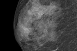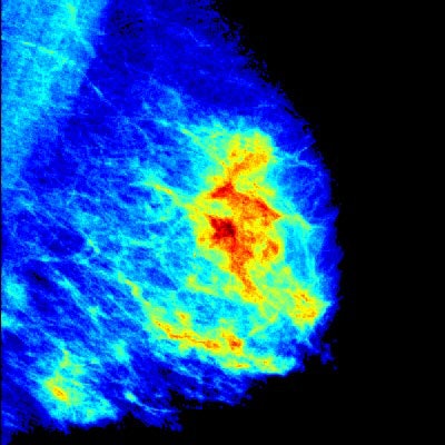
Breast imaging software can be reliable in estimating the local glandular tissue distribution in mammograms and can be used for assessment and follow-up, according to Spanish researchers. In addition, the software can handle small variations of the acquisition angle and to the beam energy.
In nearly 100 pairs of repeated mammograms in which each pair was acquired in a short time frame and a slightly changed position, the researchers evaluated spatial glandular volumetric tissue distribution and density measures using Volpara Solutions' Density Maps software. The team, led by doctoral student Eloy García from the Computer Vision and Robotics Institute at the University of Girona, found the software works effectively.
"This study could provide more reliability of density estimations within the radiologist's community, as well as opening a window to start considering local breast distribution for risk assessment," corresponding author Oliver Diaz, PhD, also from the Computer Vision and Robotics Institute, told AuntMinnieEurope.com. "We believe that local distribution of breast density can provide additional information to identify regions with high probability of malignancy."
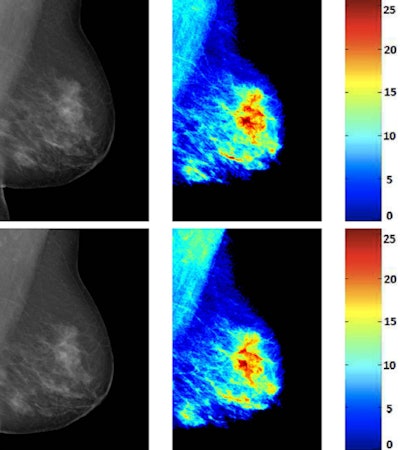 Reacquired mammograms of the same patient and their corresponding breast density maps from Volpara. The amount of breast tissue is shown in a color map along with their label in millimeters. All images courtesy of Oliver Diaz, PhD.
Why breast density?
In the quest to improve breast cancer screening, researchers are turning to a more personalized approach. Volumetric breast density, local density distribution, and parenchymal patterns have come into focus as important risk factors. Vendors such as Volpara and Hologic have developed several software tools to estimate breast density from mammograms.
"However, performing an automatic evaluation of the spatial distribution of the glandular tissue is a challenging task," the authors wrote (European Journal of Radiology, 30 May 2017). "On the one hand, few algorithms provide pixel-wise information about the breast glandularity. On the other hand, several factors, such as the breast compression or temporal changes (aging, involution, hormonal interactions) can modify the appearance of the mammogram as well as the automatic density measures."
In their study, which Volpara did not fund, they sought to evaluate the repeatability of the glandular tissue measures provided by the firm's Density Maps. The software uses a physics-based model to extract pixel-wise information from raw mammograms. Few attempts have been performed for evaluating the density maps obtained using this software, according to the researchers.
García and colleagues used repeated mammograms for quantitatively assessing the variation of the density maps. There was no change in the glandularity of the breast, although several factors, such as different breast compression or different acquisition parameters, can modify the appearance and density measures of the breast, they wrote.
In their retrospective analysis of 99 repeated mammograms acquired between 2008 and 2016, the researchers compared the density and glandular tissue between the two acquisitions. They evaluated the local glandular information using histogram similarity metrics, such as intersection and correlation, and local measures, such as statistics.
They found a high correlation (breast volume R = 0.99, volume of glandular tissue R = 0.94, and volumetric breast density R = 0.96) regardless of the anode/filter material. Also, the histogram intersection and correlation metric showed for each pair the images share a high degree of information.
"Despite the different parameters of both acquisitions, the results show an important agreement," the authors wrote. "We also compare in a global way the information contained in the density maps using histogram similarity metrics, in particular the histogram intersection and correlation. Both metrics showed a high degree of similarity between the information contained in the two density maps."
However, breast thickness affected the glandular tissue distribution, which was anticipated, and deformable registration algorithms can help mitigate the effect, they added.
The researchers are conducting a new study to compare global and local breast density measures in both full-field digital mammography and breast tomosynthesis, according to Diaz. The work is in its initial stages, but the researchers plan to have something ready for the end of the year.
"We are gathering data at the moment from our partner Hospital Parc Taulí," Diaz wrote in an email. "The idea is to obtain images from both tomosynthesis and mammography from the same patient and acquired within a short time frame. Then we will calculate both global and local density measures and will investigate how they differ from one technology to another."


