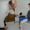
The clinical integration of 3D printing and extended reality technologies is having a growing impact on patient care by improving surgical planning and aiding simulations to guide procedures in real-time and better educate trainees, according to researchers from Croatia. The potential applications of 3D printing are limitless, they insist.
"3D printing advancements enable the production of customized surgical guides, implants, and prosthetics tailored to the unique characteristics of each patient," Dr. Antonia Ivančić, from the department of radiology at Nacionalna Memorijalna Bolnica (NMB) "Dr. Juraj Njavro" Vukovar, noted. "The ongoing progress in image processing and computational speed is moving us closer to the realization of seamless, transparent, and gesture-controlled intraoperative overlays, promising to further enhance surgical visualization."
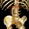
 A CAD representation (a) and 3D printed model (b, c) of the pathologically altered mandible were created for preoperative planning and osteosynthetic plate prebending. Personalized surgical guides designed for mandible reconstruction surgery with free fibula flap are shown (d, e). The fitting of the flap's bone fragment to the native mandible post plate fixation is depicted (f). A cinematic-rendering reconstruction from a follow-up CT scan, taken 13 months after the reconstruction surgery, demonstrates complete healing of the bone fragment; its edges are marked with red arrows (g). All figures courtesy of Dr. Vjekoslav Kopačin, PhD, Department of Diagnostic and Interventional Radiology, University Hospital Center Osijek, Croatia, and presented at ECR 2024.
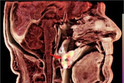
Cinematic rendering transforms standard CT and MRI data into photorealistic 3D images by precisely simulating light propagation and interaction, and it enhances depth perception in 3D reconstructions, which is crucial for through-plane views, she explained. It allows surgeons to visualize anatomical details from various perspectives, closely resembling what they encounter in the operating room.
"The increased realism may help mitigate the impact of unexpected findings and anatomical variations during surgery," stated Ivančić and coauthor Dr. Vjekoslav Kopačin, PhD, who shared their experiences at ECR 2024. "It provides a lifelike portrayal of anatomical features, offering improved depth and structure delineation. In instances where 3D printing is unavailable, it can serve as a viable alternative.
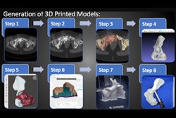
3D printing, augmented reality (AR), and virtual reality (VR) are being used as 3D imaging techniques to display anatomical models. The process involves converting original stacks of 2D radiological images (i.e., DICOM data) into a 3D volume, comprising image acquisition, segmentation, and computer-aided design/modeling. The manipulated data is then optimized for the chosen visualization method: 3D printing, AR, or VR.
"These 3D models simplify the understanding of intricate diseases," they pointed out. "The next step is immersing surgeons in a virtual 3D space with VR goggles. CAD 3D models are previously segmented and generated using software and imported into visualization applications, enabling CAD 3D model manipulation using controllers."
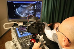
CT images are often preferred for 3D printing due to their versatility and ease of postprocessing. The notable contrast, signal-to-noise ratio, and spatial resolution improve the distinction of structures and reduce potential issues like partial volume effects that might constrain 3D printing, according to Ivančić and Kopačin. Creating anatomically precise 3D models requires meticulous planning, precise acquisition of DICOM data, specialized software for segmentation, CAD tools for manipulating 3D components, and the expertise of skilled operators.
"3D printing offers top visual and tactile anatomy experiences. Combined with 3D modeling, it allows creating patient-specific surgical guides and grafts," they noted. "Additionally, open-source technologies and free software are utilized in alternative approaches for mandibular reconstruction planning, providing cost-effective options compared to expensive proprietary software solutions."
 3D printed model of the osteolytic skull lesion in right fronto-orbital region was created for preoperative planning with 3D printed personalized surgical guide (a), 3D printed mold for personalized graft generation (b), osteotomy using the personalized surgical guide, CRT reconstruction shows impeccable fit and alignment of the molded graft.
The introduction of 3D printing improves preoperative planning, providing surgeons with a clear understanding of tumor resection extent, fibular bony fragment size and shape, and osteotomies fitting, Ivančić and Kopačin added. This enhances precision and efficiency in restoring facial contour after tumor resection, especially in deciding osteotomies.
"Effective utilization of medical 3D printing to improve patient care requires strong interdisciplinary communication, collaboration, knowledge sharing, and a deep understanding of technological advancements. The potential applications of 3D printing in the medical field are boundless and constrained only by one's creative imagination," they concluded.
You can view the researchers' full exhibit on the EPOS website of the European Society of Radiology.