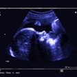
Ultrasound is a useful, dynamic tool for pediatric lung imaging, according to a presentation given February 28 at ECR 2024.
In his talk, Jovan Lovrenski, MD, PhD, from the University of Novi Sad in Serbia discussed the diverse ways ultrasound can detect abnormalities in the lungs despite skepticism surrounding the modality’s use over others.
“It [lung ultrasound] can be a very helpful diagnostic tool,” Lovrenski said.
As more complex imaging modalities emerge as ways to detect abnormalities and screen for diseases, ultrasound in pediatric imaging may seem like a simple tool that has its share of shortcomings.
Lovrenski said in the era of skepticism a few decades ago, clinicians had perceptions about ultrasound’s use that prevented its widespread use in pediatric imaging. He said a few reasons included bones and air within and around the lungs being considered an insurmountable obstacle, pathologies that border the pleura, and clinicians not being used to using the modality.
However, technological advancements and studies over the decades have made way for lung ultrasound’s use in pediatric imaging. Lovrenski said this era of enthusiasm began in the ‘90s and that the subspecialty is still in this era.
The main reason for using lung ultrasound in children is to detect pneumonia and neonatal lung diseases. Meta-analyses Lovrenski cited showed that lung ultrasound has higher sensitivity than that of chest x-ray, as well as comparable specificity.
“Behind a simple chest x-ray, there’s a whole spectrum of potential findings that lung ultrasound can reveal to you,” he said.
Lovrenski also highlighted that lung ultrasound can also detect areas of necrosis smaller than 5 mm and that the modality can be used for frequent follow-up imaging without exposing pediatric patients to potentially harmful effects. Examples include multiple follow-up exams within days to track pneumonia progression and areas of necrosis.
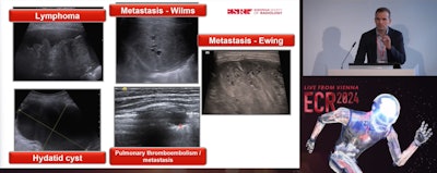 Jovan Lovrenski, MD, PhD, from the University of Novi Sad in Serbia explains the diverse use of lung ultrasound in pediatric imaging in an ECR 2024 presentation.
For neonatal lung diseases, Lovrenski said one crucial point of ultrasound use is correlation with clinical and lab findings. This is because lung ultrasound findings can change based on different stages and grades of the same disease.
Functionally speaking, ultrasound can also be used to predict the need for surfactant replacement therapy and intubation and the development of bronchopulmonary dysplasia (BPD).
“In all studies, it’s proven that lung ultrasound can truly help clinicians make more targeted therapeutic choices, especially in neonatal intensive care units (NICUs),” Lovrenski said.
However, Lovrenski also cautioned audience members about over-enthusiasm on lung ultrasound’s use in pediatric imaging. He cited a 2020 study that suggested the modality can completely replace chest x-ray for diagnosing neonatal lung disease and reasoned that there is no perfect diagnostic modality.
“Going too far with the promotion of lung ultrasound might unfortunately compromise all its benefits and valuable advantages,” he said. “At the end, there is a thin line between success and failure, and we don’t want this to jeopardize all the fantastic things that lung ultrasound brings to our everyday clinical practice just by overusing it.”


