The RSNA spotlight fell on Peruvian radiology on 3 December, when a special session focused on the role of imaging in pneumoconiosis and pediatric brain parasitic infections and in the investigation of ancient mummies.
"Pneumoconiosis is a diagnosis that affects not only the patient but the physician as well," noted Dr. Luis Antonio Campos Calderon, president and CEO of the Instituto de Imagenes Medicas S.A.C. in Lima. "Proper training and clinical history is always needed. Proper workflow and laws are needed to avoid legal issues. Increasing awareness and education of the population at risk is a must."
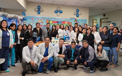 Staff at the Department of Diagnostic Imaging at Instituto Nacional de Salud del Niño de San Borja in Lima, Peru, where RSNA is establishing a Global Learning Center. Photo courtesy of RSNA.
The most common types of pneumoconiosis are asbestosis, silicosis, and coal miner’s lung. These particles cause inflammation and fibrosis in the lung, resulting in irreversible lung disease, he pointed out. Occupational inorganic and coal dust exposures can be broadly categorized as strongly fibrogenic dusts (silica, asbestos, and coal), in addition to other inorganic dusts and metal fumes that may be associated with adverse respiratory effects (e.g., iron, emery, graphite, gypsum, marble, mica, perlite, plaster, silicon, silicon carbide, soapstone, talc, and welding fume particles).
In these cases, it's important to be aware of what the usual workflow looks like as well as well as what's missing and what legal caveats we may find, Campos said. It's vital to describe the reality of the pneumoconiosis diagnosis in Peru, assess the implications for physicians and patients, and present a range of cases in different settings.
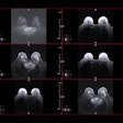
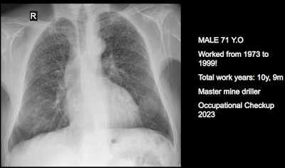
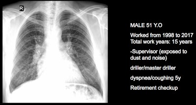 Three clinical cases courtesy of Dr. Luis Antonio Campos Calderon.
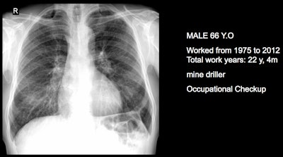
The International Labor Organization (ILO) International Classification of Radiographs of Pneumoconioses is used for epidemiological studies, screening, and surveillance of workers exposed to dust in the workplace, and clinical purposes, and the National Institute for Occupational Safety and Health (NIOSH) B Reader Program certifies physicians in the ILO classification system. Each case should discuss its ILO classification and brief comments about the workflow, he noted.
"Pneumoconiosis often develops slowly, and symptoms may not appear until significant damage has occurred. Early detection is crucial but can be difficult due to limited resources and awareness," Campos explained. "This diagnosis can be fraught because in Peru workers diagnosed with pneumoconiosis have rights to compensation and healthcare under current labor laws and social security systems. However, navigating the legal and bureaucratic processes to claim these benefits can be complex and challenging."
The main diagnostic criteria are: Work history (minimum 10 years exposure to silica dust or other particles); chest x-ray (micronodular pulmonary fibrosis, coded according to ILO classification); progression (image profusion increases over time, even after exposure cessation; irreversibility (disease is progressive and irreversible, no stable pneumoconiosis over time).
The incidence of pneumoconiosis is generally higher among those with a history of working around coal, asbestos, or silica. A 2016 study found that a lack of education, residing in the Peruvian highlands, and working underground for over 20 years are associated risk factors.
"This topic has been chosen because Peru is a mining country, and we are also opening deep megaports so ship cleaning with sand may increase the cases if not managed correctly," Campos told AuntMinnieEurope.com ahead of the RSNA session.
Impact of geography and socioeconomics
Radiology in Peru is shaped by the country's geography, healthcare infrastructure, and socioeconomic conditions, and there is a growing emphasis on teleradiology to improve access and quality of care across the country, according to Dr. Luis Alberto Chávez Saavedra, president of the Sociedad Peruana de Radiologia (SOCPR).
"Peruvian epidemiology is not only related to complex cases of infectious diseases, but also a significant number of cancer diagnoses, hereditary conditions, and metabolic disorders, much like those encountered in the developed world," he said, adding there are currently around 1,500 radiologists in Peru serving a population of about 34.3 million. A total of 567 live in Lima, and over 550 people attended the 28th Peruvian Congress of Radiology, held from 3 to 5 October 2024 in Lima.
Raising awareness of pediatric brain parasitic infections is essential because these conditions carry high rates of morbidity and mortality, stated Dr. Carlos F. Ugas Chacape, a Peruvian pediatric radiologist and former president of the Latin America Society of Pediatric Radiology (SLARP) who currently is head of the Pediatric Radiology Section of UH Rainbow Babies & Children's Hospital in Cleveland, Ohio.
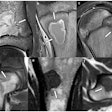
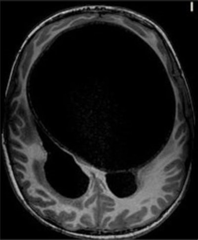 An axial T1-weighted MRI scan of a hydatid cyst. Clinical images courtesy of Dr. Carlos F. Ugas Chacape.
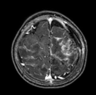 Contrast-enhanced T1-weighted MRI scan shows multiple abscesses due to free-living amebiasis (Balamuthia mandrillaris).
Neurocysticercosis is still prevalent but is decreasing in Peru, whereas free-living amebiasis is less common but has a rising prevalence and is often fatal without treatment. Toxoplasmosis remains a significant congenital infection in Latin America, and it's important to be aware of hydatid disease, malaria, toxocariasis, and strongyloidiasis.
"Parasitic brain infections persist in Peru, and primarily affect underserved children with poor access to medical care," Ugas said. "Neurocysticercosis is still relevant, and the radiological features and stages in children align with adults."
Free-living amebiasis is a growing concern, being rapidly progressive and often fatal without early intervention. Congenital toxoplasmosis remains prevalent, and the severe features are more prevalent in Latin America.
Radiologists play a vital role by making a timely diagnosis using MRI, improving outcomes, and guiding treatment. "Our call to action should be advocacy for improved sanitation, healthcare access, and education to combat these infections," he said.
The RSNA session also included a talk on the imaging used on the Lady of Cao, a mummy found in the Moche culture in Northern Peru. The unique aspect of this mummy is that she may have been a high-ranking priestess or even a Moche ruler. Prior to this discovery, it was believed that only men held high rank in the Moche culture.
"Applying medical radiology techniques in a historical and cultural context would allow us not only to preserve heritage but also to obtain valuable information about funerary practices and the health of ancient civilizations," said presenter Dr. Pedro Tapia.
More details on the session are available here.
For our full coverage of this year’s meeting, visit our RADCast.


