An ultralow dose imaging method used with long axial field-of-view PET scanners produces images with a radiopharmaceutical dose 50 times lower than the standard effective dose, according to research presented at the Society of Nuclear Medicine and Molecular Imaging annual meeting in Toronto.
The technique can generate quantitative PET images without the need for accompanying CT scans, which cuts patients' radiation dose exposure, noted lead author Hasan Sari, PhD, of Bern University Hospital and Siemens Healthineers International in Switzerland.
"This reduction in radiation dose is 50 times lower than the standard PET dose and is comparable to the dose received from a mammogram or a chest CT radiograph," he said in a statement released by the SNMMI.
PET/CT scans expose patients to radiation by both the dose that results from the administered radiopharmaceutical and from the CT scan itself. Although the use of PET scanners has reduced this radiation exposure, this effect has been hindered by the radiation dose required for the CT scans conducted for PET attenuation correction, Sari and colleagues noted.
"Recently, methods have been developed to utilize the background radiation from Lutetium-176 that is found in some PET scintillators as transmission source; this is known as LSO-TX," he said in the statement. "By combining LSO-TX information with joint reconstruction- and deep learning-based methodologies, CT-free attenuation maps can be generated."
The investigators conducted a small study with four patients who received 7.9 MBq of radiotracer and underwent a 90-minute PET scan using a long axial field-of-view PET scanner; they also underwent low-dose CT (LDCT) imaging. They then generated CT-based and LSO-transmission-based attenuation maps to reconstruct PET images and compared these across a variety of scan lengths.
The group observed close visual resemblance between CT- and LSO-TX-based attenuation maps and between PET images reconstructed with the maps. By using the LSO-TX-based attenuation correction alone (that is, no CT imaging), the total effective dose in these scans was reduced to approximately 0.15 mSV while "still preserving good quantitative accuracy and maintaining clinically feasible scan durations."
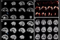
 Top row: CT- and LSO-TX-based attenuation maps with their respective percentage relative change (%RC) map. Bottom row: PET images reconstructed using CT- and LSO-TX-based attenuation maps with their respective %RC map. Image and caption courtesy of the SNMMI.
"Ultralow-dose protocols have the potential to extend the use of PET scans beyond their current applications and could significantly enhance the utility of this modality in screening studies involving at-risk or healthy subjects, research studies, enhanced treatment response assessments with more frequent follow-up scans, and pediatric scans," Sari concluded.
Click here for our full SNMMI coverage.