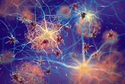
The winner of this year’s Image of the Year award at the annual meeting of the Society of Nuclear Medicine and Molecular Imaging (SNMMI) in Toronto reveals brain nuclei activity with a dedicated brain PET scanner.
The image was chosen from more than 1,500 abstracts submitted for the congress. It comes from a study using what the researchers have named NeuroExplorer, or “NX,” an ultrahigh-performance PET system with 10 times the sensitivity and resolution over current high-resolution research tomograph (HRRT) systems, according to its developers.
Principal investigator on the project, Richard Carson, PhD, director of the PET Center at Yale University in New Haven, Connecticut, U.S., said the technology has the potential to spur advances in the treatment of many brain diseases.
 F-18 SynVesT-1 images at early (0-10) and late (90-120 minute) times postinjection. There is clear identification of high flow regions in the early images. The late images show the synaptic (SV2A) pattern which differs from the flow pattern, e.g., in the thalamus. B. 11C-PHNO binding potential (BPND) images shown in transverse, coronal, and sagittal orientations of PET alone and PET overlaid with MRI. Left: Region of substantia nigra (green arrow, max display: 4.0). Right: Thalamic region (max: 2.5) showing focal bilateral binding in a specific thalamic nucleus (blue arrow, likely anteroventral nucleus). C. Sagittal images of 11C-LSN3172176 M1 muscarinic cholinergic BPND (max display: 10). D. R1 images of the same tracer (max: 2). Cerebellum (blue arrow) shows no specific binding (C) and high tracer delivery (D). E. 18F-FE-PE2I dopamine transporter BPND images (zoomed, max display: 6) showing striatum and substantia nigra (green arrow). F. R1 images of the same tracer (max: 2) with inset showing zoomed region in E.Image courtesy of Richard Carson, PhD
“The high resolution of NeuroEXPLORER images is due to the system’s unique detector design and exceptional sensitivity produced by its long axial field-of-view. This technology will provide the opportunity for advanced research on all types of neuronal molecular and functional activity,” he said, in a news release.
In the study, Carson and colleagues conducted human brain imaging with both the NeuroEXPLORER and the latest HRRT system. Multiple PET radiotracers were administered to observe synaptic density, dopamine receptors and transporters, muscarinic cholinergic receptors, and glutamate receptors. Images from both scanners were then compared.
A striking improvement in image contrast and quality of the NeuroEXPLORER compared to the HRRT was evident, according to the findings. The NeuroEXPLORER images demonstrated low noise and exquisite resolution, showing focal uptake in specific brain nuclei.
While the NeuroEXPLORER is currently used for research purposes, Carson said he hopes that once the excellent image quality is recognized by physicians it will become available for clinical use.
Each year, SNMMI chooses an image that best exemplifies the most promising advances in the field of nuclear medicine and molecular imaging. The state-of-the-art technologies captured in these images demonstrate the capacity to improve patient care by detecting disease, aiding diagnosis, improving clinical confidence, and providing a means of selecting appropriate treatments, SNMMI said.
“The dramatic improvement in resolution and overall quality of the NeuroEXPLORER images compared to the HRRT images is clear,” noted SNMMI Scientific Program Committee Chair Dr. Heather Jacene in a statement. “The NeuroEXPLORER has the potential to be a game changer in research for conditions such as Alzheimer’s disease, Parkinson’s disease, epilepsy, and mental illnesses.”
The NeuroEXPLORER scanner was built as a collaboration with Yale University; University of California, Davis; and United Imaging Healthcare of America, and it was funded by a National Institutes of Health Brain Initiative grant.
Click here for our full SNMMI coverage.