Sarah Bohndiek, PhD, and her colleagues at the University of Cambridge in the U.K., are on a mission to develop low-cost and nonionizing optical technology to improve early cancer detection. Bohndiek heads up an international team combining physicists, engineers, chemists, and molecular biologists who work at the clinical interface.
"My lab is split between the department of physics and the Cancer Research UK (CRUK) Cambridge Institute, which serves as a pipeline that allows us to develop novel hardware and data analytics, and then bring those early technologies into preclinical trials in the institute," she said.
The team also benefits from strong links with the local hospital -- Addenbrookes -- which is just a short walk from the institute. Thanks to the partnership, scientists and clinicians can exchange ideas and discuss how new technologies could fit into and streamline cancer diagnosis.
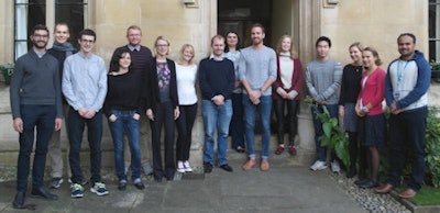 Sarah Bohndiek, PhD, and her research group at the University of Cambridge. All images courtesy of the University of Cambridge.
"We're working with Rebecca Fitzgerald and Massimiliano di Pietro, who are gastroenterologists," Bohndiek explains. "They are performing endoscopies on patients on a regular basis, and can direct us to the particular challenges, which helps to guide our research."
One of Bohndiek's major project goals is to improve the visibility of early cancer in the esophagus. In many cases, cancer that starts in this part of the body is preceded by a condition known as Barrett's esophagus.
"This condition often arises when people have chronic reflux, and it involves a change in the lining of the esophageal tissue," Bohndiek said. "Patients with Barrett's have about a 0.5% per year risk of going on to develop cancer, but as soon as abnormal lesions known as dysplasia appear, their risk rises to around 10% and even up to 30%."
As part of the monitoring program, gastroenterologists use a camera to image the esophageal tissue -- and it's here the Cambridge team is applying its talents.
"Currently, the contrast that allows the gastroenterologist to pick out early lesions is really poor," Bohndiek said. "The precancer can look very similar to the background Barrett's tissue, or the background healthy tissue."
Multidimensional imaging opportunities
Diagnosing dysplasia at an early stage allows clinicians to treat the condition endoscopically to prevent cancer progression. And by applying a combination of hyperspectral and holographic imaging, the researchers plan to augment the vision of the gastroenterologist and make the dysplasia much more visible.
"If you can sample colors on a much finer scale, you boost your chances of picking out biological changes that give rise to cancer," Bohndiek said. "Additionally, by considering the angle at which the light travels you may be able to gain further contrast between healthy and unhealthy tissue, as it is sensitive to order and disorder."
In the lab, and together with its collaborators, the group is developing detectors that can sample over many more channels than the red, green, and blue frequencies typically found in normal cameras.
Today, hyperspectral imaging is used by the military to monitor the movement of troops and equipment, with apparatus deployed on unmanned aerial vehicles. But to be fit for medical use, Bohndiek and her coworkers must find a way to improve the imaging speed of the apparatus and make it sufficiently robust to withstand service on a hospital ward.
"In a clinical setting, these instruments will have to be able to be wheeled around on trolleys and resist knocks and bumps, while maintaining their alignment," Bohndiek said. "The idea behind our approach is to create the hyperspectral and the holographic components initially as two separate instruments and then ultimately combine them."
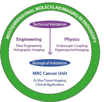 Physicists, engineers, chemists, and molecular biologists collaborate on the multidimensional molecular imaging project.
Twelve months into the three-year project -- funded by the CRUK-EPSRC (Engineering and Physical Sciences Research Council) project award that supports the formation of multidisciplinary cancer research teams -- the team already has prototypes up and running on the bench top. Bohndiek's research partners on the project also include Tim Wilkinson from the department of engineering, who works on telecommunications and optical fibers and brings experience in modal propagation of light. It's another key collaboration that allows the scientists to pool their knowledge to advance endoscopic imaging.
Beyond enhancing the detection of esophageal cancer, the outcomes of the project have the potential to improve the early diagnosis of other cancers of the gastrointestinal tract and lung. And the researchers are mindful of the benefits of sharing knowledge to broaden the application of their work.
"Rebecca [Fitzgerald] and I run an interdisciplinary program focused on early detection, as part of our Cambridge Cancer Centre activities," Bohndiek said. The center hosts regular networking events and breakfast meetings where physical scientists and engineers are invited to speak with clinicians to discuss opportunities for applying new technologies and techniques. "The goal is to stimulate research at the borders of the various scientific disciplines, and develop initiatives to improve communication and help people build up a greater number of interdisciplinary projects at the university."
Editor's note: This article is the first in a series looking at some of the research funded through the CRUK-EPSRC Multidisciplinary Project Award. In addition to this scheme, Cancer Research UK offers a range of funding opportunities open to researchers not currently working in cancer who are seeking to focus their expertise to this area: Pioneer Award; Early Detection Programme and Project Awards; and Grand Challenge.
© IOP Publishing Limited. Republished with permission from medicalphysicsweb, a community website covering fundamental research and emerging technologies in medical imaging and radiation therapy.


