As molecular imaging continues to develop, it's worth considering the origins of nuclear medicine, which has a long and rich history. Appreciating the early days of nuclear medicine can lead to a better understanding of today's developments.
It was physicist Max Born who told the tale of the typesetter who printed "unclear science" instead of "nuclear science." I have also heard of "i-suppose-o-topes" instead of "isotopes," when referring to imprecise nuclear medicine images and reports. Modern nuclear images are anything but imprecise, however, and are leading to an ever deeper awareness of physiological and pathological processes in disease.
Developments since the early years have been prodigious and include PET/CT, PET/MRI, and SPECT/CT, using ever more sophisticated radiopharmaceuticals. Yet the basic principles established by the pioneers remain the same and are still relevant.
Although atoms were proposed by Democritus more than 2,000 years ago, it was Robert Boyle (1627-1691), the first atomic scientist, who realized that there were many different types of atoms, or first principles, of which matter is composed. John Dalton (1766-1844) took further steps forward and proposed that atoms are separate real material particles that cannot be subdivided. He also stated that atoms of the same element are similar in all respects and equal in weight, atoms of different elements have different properties, and "compounds" are formed by the union of atoms of different elements in simple numerical proportions.
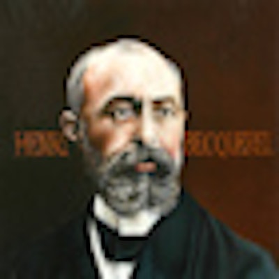
 Henri Becquerel
All this was to change for classical physics when x-rays were discovered by Wilhelm Röntgen in 1895, closely followed by Henri Becquerel and his Becquerel rays in 1896. Whilst the medical imaging applications of x-rays were immediately realized, the use of radioactive materials took considerably longer to implement, apart from the use of radium for therapy. This was because apparatus for radiography was readily available throughout the world, and the techniques needed were relatively simple.
Radioactivity was an entirely different matter, and it was very difficult to collect a significant amount of radioactive material to be useful. Marie Curie coined the term "radioactivity," and she spent many years with her husband Pierre in isolating and defining naturally occurring radioelements. Marie Curie's daughter, Irène, and her husband, Frederick Joliot, discovered artificial radioactivity in 1934 when they were creating radioactive nitrogen from boron, as well as radioactive isotopes of phosphorus from aluminum, and silicon from magnesium. For this achievement, they were awarded the Nobel Prize for chemistry in 1935.
It was only following the development of the cyclotron and nuclear reactors that radioisotopes with a medical use (other than for therapy) could be produced. The cyclotron was invented in 1931 by Californian Ernest Lawrence, and the first cyclotron for biomedical research was opened in 1940 in Cambridge, MA. The original nuclear reactor was first made to work successfully in a squash rackets court under a sports stadium in Chicago on 2 December 1942.
 George Hevesy
The use of radioactive material as a biological tracer was first described by the Hungarian George Hevesy (1885-1966), who received the Nobel Prize in 1943. In 1923 Hevesy had codiscovered hafnium. There was a perceived gap in Mendeleev's periodic table for an element with 72 protons. Using a sample from the mineralogical museum in Copenhagen, Hevesy found a characteristic x-ray spectral recording, proving the presence of the new element. This jointly earned him the 1943 Nobel Prize in chemistry with Dirk Coster.
Hevesy had worked with Ernest Rutherford on radium D, an isotope of lead. In experiments in 1913, Hevesy mixed a known amount of pure radium D with a known amount of a lead salt and followed up "the paths of the lead atoms using radium D ... as an indicator." Using a naturally occurring radioisotope of lead, Hevesy studied the distribution of lead in the broad bean plant in a series of classic experiments. This is the tracer principle, allowing us to follow molecules in chemical reactions, and as Dr. Henry Wagner, the U.S. nuclear medicine pioneer, said, "It is as if a molecule emitted a radiosignal, telling us what it was doing at all times." This is the basis of modern nuclear medicine and molecular imaging.
The first tracer studies in humans were carried out by Hemann Blumgart and co-workers in Boston in 1925. Blumgart injected a solution of radium C into a patient's right arm and measured the time it took for the radioactivity to be detectable in the left arm, using a Wilson cloud chamber. This measured the circulation time and was measurable and independent of the amount of radioisotope injected.
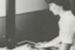
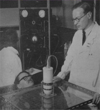 Keith Halnan performs external counting with a scintillation counter in 1957.
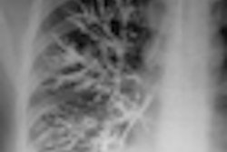
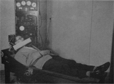 Measuring thyroid activity with radioiodine, from Keith Halnan (1957).
The initial clinical work in the 1940s using reactor-produced radioisotopes was primarily related to the thyroid using radioactive iodine (I-131). The original work involved using a Geiger-Müller detector placed at different marked anatomical points over the neck and upper chest. This progressed to automatic (rectilinear) scanning in the early 1950s and the gamma camera, invented by Hal Anger in the late 1950s.
Dr. Adrian Thomas is chairman of the International Society for the History of Radiology and honorary librarian at the British Institute of Radiology.
The comments and observations expressed herein do not necessarily reflect the opinions of AuntMinnieEurope.com, nor should they be construed as an endorsement or admonishment of any particular vendor, analyst, industry consultant, or consulting group.
Further reading
- Halnan KE. Atomic Energy in Medicine. London: Butterworths Scientific Publications; 1957.
- Mallard J, Trott NG. Some aspects of the history of nuclear medicine in the United Kingdom. Seminars in Nuclear Medicine. 1979;9(3):203-217.


