Priority should be given in pediatric imaging to CT scans of specific body areas at risk instead of full-body CT, researchers from Denmark have asserted. Also, better education of the trauma team and flow charts indicating when to scan in children may also help to reduce overall radiation dose, they say.
"Full body trauma CT exposes the patient to high levels of ionizing radiation, meaning that scanning must be appropriate and justified," noted Dr. Simona Gentile, from Sjællands Universitetshospital in Roskilde and colleagues at Copenhagen University Hospitals. "Using the ALARA (As Low As Reasonably Achievable) principle, image quality should not be compromised -- specific, optimized CT protocols on the scanner for different age groups should be available. Software in the scanners may help reduce radiation to sensitive tissue such as the breast and should be activated."
For all trauma patients aged under 18 who underwent CT at Rigshospitalet, Copenhagen University Hospital, between 1 January 2019 and 31 December 2022, the team decided to map which body areas were scanned and to evaluate radiation doses with relation to the injury mechanism.
The follow-up period was one month from the date of arrival, and the researchers defined having an injury in a body area as an abbreviated injury score (AIS) severity ≥ 2. The score can be between 1 and 6, where 6 is fatal. They compared radiation doses of CT thorax-abdomen to the dose length product (DLP) of CT abdomen exams, together with the DLP of CT thorax exams from the European diagnostic reference levels (DRLs).
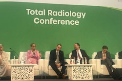
Gentile and colleagues presented their results at EuroSafe 2024. A total of 544 patients were included in the full cohort, of which 429 (79%) were primary admissions to the major trauma unit at Rigshospitalet, a tertiary referral center. The median age was 11, and 35% were girls. The three most common injuries were motor vehicle accident (34%), fall from a height (33%), and struck/hit by blunt object (12%).
A total of 50 patients (12%) did not undergo CT. Among all 429 patients, full body trauma CT was performed in 258 patients (60%), CT of the brain and cervical spine in 45 (10%), CT of the thorax-abdomen in 34 (8%), brain CT in 22 (5%), CT of the abdomen in 5 (1.2%), while 15 patients underwent other combinations (e.g. CT of the cervical spine and thorax).
Among patients involved in motor vehicle accidents who had a full-body trauma CT scan, 40 (31%) did not present with any injury, while 56 (43%) were injured in only one body region and 33 (26%) were injured in two or more regions. The same trend was seen for the other injury mechanisms.
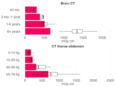 Ionizing radiation doses for brain CT and thoracic-abdominal CT. Ruby bars: European DRLs. Box and whisker plots: quartiles and median of doses in the included patients. Figure courtesy of Dr. Simona Gentile et al and EuroSafe 2024.
Compared with the European DRLs, the radiation doses for brain CT were not significantly different from the reference level in the age group 1-6 years (p = 0.956), while doses were significantly higher in the oldest age group (p < 0.0001). This might be due to the selective trauma population presented in the study and the frequent use of supplementary facial bone CT.
The values for CT thorax-abdomen were slightly (yet significantly) lower both for the weight groups 5-14 kg (p < 0.0001) and 15-29 kg (p = 0.0008). The older patients received slightly (yet significantly) higher doses than the reference levels with p < 0.0001 for the weight group 30-49 kg and p < 0.010 for the weight group 50-79 kg. This was in part due to some patients having supplemental contrast phases, possible cervical CT angiography dose registered under the CT thorax-abdomen, and some patients having a body composition with large muscles or being obese. Also, the general quality of the trauma protocols was higher than other follow-up CTs.
"The strengths of our study include large population covering four years, recent data as well as reliable data from our local trauma register used for quality assurance," the authors stated. "Our study also has limitations, such as few included patients in the youngest age group and area-specific doses unavailable in some patients."
The co-authors of the EuroSafe presentation were Dr. Lotte Borgwardt, Dr. Lasse Høgh Andersen, Dr. Oscar Rosenkrantz, Dr. Søren Steemann Rudolph, and Dr. Caroline Ewertsen. You can access the full exhibit on the EPOS website of the European Society of Radiology.


