
Prof. Gianfranco Gualdi, the senior radiologist from Rome who faces manslaughter charges over alleged malpractice, gave a presentation at a meeting held on 14 November. A video clip of him has appeared on social media.
Along with two other radiologists and a cardiologist, Gualdi is being prosecuted over the death of the well-known investigative journalist Andrea Purgatori. An initial hearing took place on 19 September, and the trial is due to begin on 20 February 2026. There has been some speculation in Italy that Gualdi may be too frail and elderly to take the stand and present his perspective in court.
 Prof. Gianfranco Gualdi summarized his presentation in an Instagram reel posted on 14 November by INI Grottaferrata, a private facility located 25 km south-west of Rome and accredited by the Italian health service.
According to an article posted by In Sanitas on 16 November, Gualdi gave an in-depth presentation at last Friday’s meeting, “Update on gastric and esophageal cancer.”
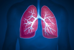
His talk was about the multimodal radiological staging of esophageal and gastric cancers, highlighting how the integration of various imaging methods has been a crucial step in improving diagnosis, treatment planning, and patient outcomes. He explained that the combination of endoscopic ultrasound, CT, MRI, and PET/CT has allowed for an increasingly comprehensive assessment of the three key elements of the TNM classification: tumor wall extension (T), lymph node involvement (N), and the presence of metastases (M).
“Multimodal staging has represented a quantum leap in the management of esophagogastric cancers,” Gualdi said. “The integration of various imaging techniques has allowed us to accurately define the extent of the disease, improve the selection of patients eligible for surgery, and increase the effectiveness of treatment protocols.”
The gold standard
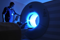
CT has established itself as the gold standard for systemic staging and surgical planning, and it can identify advanced lesions with high accuracy and provide essential information on lymph node and metastatic involvement, he explained. Thanks to modern multiphasic techniques, CT has enabled the recognition of peritoneal or hepatic invasion and the differentiation of the different stages of the disease, which are crucial elements for defining the therapeutic pathway, In Sanitas reported.
MRI has made a crucial contribution to tissue characterization and the evaluation of liver and peritoneal metastases due to the high sensitivity of diffusion-weighted sequences and the use of liver-specific contrast agents, Gualdi continued. In esophageal cancer, T2-weighted and diffusion-weighted sequences have allowed radiologists to distinguish superficial tumors (T1-T2) from deeper ones (T3-T4), achieving sensitivities and specificities comparable to or superior to those of CT, he added.
The integrated approach, involving ultrasound, CT, MRI, and PET/CT, has enabled a more precise and personalized assessment of cases, laying the foundation for increasingly targeted oncologic medicine capable of adapting treatment to the biological and morphological characteristics of each individual patient, he said.
Gualdi is thought to have been retired since 2019. His former colleagues at La Sapienza University Hospital in Rome -- radiologists Dr. Claudio Di Biasi and Dr. Maria Chiara Colaiacomo and cardiologist Dr. Guido Laudani -- are also facing manslaughter charges. Their roles in the diagnostic process and the subsequent treatment decisions of Purgatori look set to come under close scrutiny at next year's trial.
To see the video clip on Instagram, click here. To watch it on Facebook, click here.