Whole-body SPECT/CT bone scan times to detect metastatic tumors in prostate cancer patients could be cut by half with the use of faster acquisition techniques, according to a study published on 12 December in EJNMMI Physics.
A team of researchers led by Dr. Samuli Arvola of Turku University in Finland evaluated the use of a shorter SPECT acquisition protocol on the diagnostic performance of whole-body technetium-99m hydroxymethylene diphosphonate (Tc-99m HMDP) SPECT/CT in prostate cancer patients. The technique reduced total acquisition time from 40 minutes to as low as 16 minutes without any loss of diagnostic performance, they found.
Such shorter acquisition times could help clinicians potentially increase the use of accurate whole-body SPECT imaging in these patients, the authors suggest.
"Whole-body Tc-99m HMDP-SPECT/CT can be acquired using a general-purpose CZT system in less than 20 minutes without any loss in diagnostic performance in metastasis staging of high-risk prostate cancer patients," the group wrote.
Whole-body bone SPECT/CT is a more accurate method than standard planar bone scintigraphy and CT for detecting bone metastases in cancer patients, yet its clinical use is limited partly due to the lack of fast acquisition protocols, according to the authors.
Current recommendations suggest an acquisition time for whole-body SPECT/CT of at least 40 minutes, but this guidance was written prior to the development of general-purpose digital cadmium-zinc-telluride (CZT) systems, which allow optimization of acquisition protocols, including imaging time, they added.
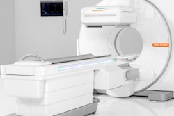
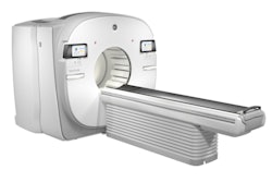
In this study, Arvola's team explored whether a Discovery NM/CT 670 CZT system (GE Healthcare) installed at Turku University Hospital could be used to cut acquisition times in a group of patients with prostate cancer, a type with high rates of metastatic bone tumors.
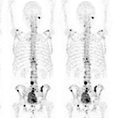
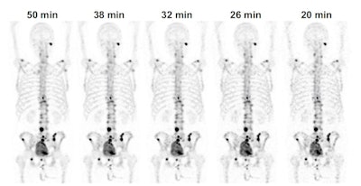 Whole-body Tc-99m HMDP-SPECT maximum intensity projections of an 80-year-old prostate cancer patient with different acquisition times. The images are filtered using a Gaussian filter with a 7-mm full-width at half maximum (FWHM) spatial resolution. Image and caption courtesy of EJNMMI Physics, licensed under CC BY 4.0 International License.
The group studied imaging from 30 prostate cancer patients who previously underwent 50-minute Tc-99m HMDP-SPECT/CT scans from the top of the head to the mid-thigh. Fifteen patients had bone metastases, and the other 15 had only benign findings. The acquired images were then resampled to produce data sets with shorter acquisition times of 41, 38, 32, 26, 20, and 16 minutes. Experienced nuclear medicine physicians qualitatively and quantitatively evaluated the images.
The investigators found that the originally acquired images had the best qualitative image quality and lowest noise. But reduced acquisition time had no effect on the diagnostic performance of the physician readers: Sensitivity, specificity, and accuracy for identifying cancer targets did not significantly differ between the 50-minute and reduced acquisition time images.
Ultimately, acquisition time is an important factor regarding the feasibility of whole-body bone SPECT/CT for imaging bone metastases. With shorter acquisition times, use could increase significantly, the authors wrote.
"[Our study validates the feasibility of] fast whole-body bone SPECT/CT in prostate cancer patients," Arvola and colleagues concluded.


