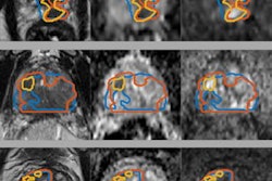
A new type of MRI protocol called synthetic correlated diffusion imaging better visualizes cancerous tissue in the prostate and may help clinicians better identify and track cancer over time, according to a study published on 1 March in Scientific Reports.
Researchers from the University of Waterloo in Ontario, Canada, developed the protocol, which captures, synthesizes, and mixes MRI signals at various pulse strengths and timings, according to a statement released by the university on 21 March. The group tested the technology on 200 patients with prostate cancer and found that it was better than conventional MRI imaging for identifying cancerous tissue.
"Prostate cancer is the second most common cancer in men worldwide and the most frequently diagnosed cancer among men in more developed countries," said lead author Alexander Wong, PhD, in the statement. "That's why we targeted it first in our research. [But] we also have very promising results for breast cancer screening, detection, and treatment planning. This could be a game-changer for many kinds of cancer imaging and clinical decision support."


