Sometimes weighing only 300 grams and suffering from multiple health complications, premature babies start life with many risks. Specialist pediatric radiologists can help identify difficulties quickly, permitting appropriate treatment. In this Q&A interview with the German Röntgen Society (Deutsche Röntgengesellschaft, DRG), pediatric radiologist Dr. Stephanie Spieth from the Carl Gustav Carus University Hospital in Dresden discusses how best to approach imaging of premature babies.
Q: Many premature babies have health complications due to their sometimes extreme immaturity. What do you come across most often as a pediatric radiologist?
 Dr. Stephanie Spieth. Photo courtesy of Carl Gustav Carus University Hospital.
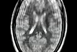
A: The immature organ systems are particularly susceptible to many diseases. In pediatric radiology, we primarily see complications affecting the lungs, abdomen, and central nervous system. Typical lung diseases initially include respiratory distress syndrome and later on, bronchopulmonary dysplasia, a chronic lung disease. In the abdomen, necrotizing enterocolitis, a severe inflammation of the intestinal wall, is particularly worrisome. In the head, oxygen deprivation and bleeding are responsible for the most common complications. These include damage to the white matter of the brain such as periventricular leukomalacia, hemorrhage, or congestion of blood around the cerebral ventricles. The hemorrhages can lead to a disturbance of the cerebrospinal fluid flow and thus to an increase in intracranial pressure, a hydrocephalus. Neurological development may be impaired in the course of disease.
Q: How can radiology help in each case?
A: Imaging can be used to make the diagnosis at an early stage, to assess the severity and to evaluate the need for intervention. In the case of premature infants, ultrasound is primarily used for cranial and abdominal examinations and x-rays for thoracic and abdominal examinations; MRI in the cranial region is used less frequently.
Q: Examining such small patients is a great challenge. What points do you have to pay particular attention to as a pediatric radiologist?
A: Strict indications are important for us, meaning that there must be a critical situation that requires further clarification. This is because every examination means stress for the premature baby which poses certain risks, for example through dislodging foreign materials, such as catheters, due to repositioning, and heat loss. This can be countered by examination in the incubator, among other things. So far, ultrasound and x-ray examinations are performed at the bedside; in the future, MRI examination will also be possible. Until then, transport incubators and in some places MR incubators are used to maintain the baby at a constant temperature.
Q: Does the examination of premature infants require special knowledge?
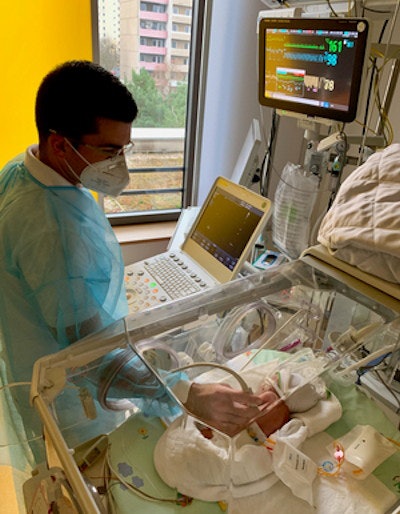
 Ultrasound exam in the incubator. Courtesy of Dr. Paul-Christian Krüger and Prof. Dr. Hans-Joachim Mentzel, Jena University Hospital.
A: Absolutely, both on the part of the technicians and on the part of the physicians, so that the examination can be performed appropriately and interpreted correctly. From a technical point of view, radiation protection is the main issue in x-ray examinations, while the small patient size is a challenge in MRI examinations. For example, a premature baby born at 24 weeks is only about 30 cm long. Moreover, in premature infants, we are not only dealing with different anatomical or physiological conditions but also with preterm-specific clinical pictures.
Q: Why is radiation protection so important in x-ray examinations?
A: Because of the particularly high radiation sensitivity of young children compared to adults, due in part to differences in anatomy and physiology, the use of ionizing radiation must be done very carefully. In addition, the lower the gestational age of the baby, the more imaging is required.
Q: Are there also safety aspects to be considered for ultrasound and MRI?
A: Intraclinical transportation with the aforementioned risks of stress due to repositioning or heat loss, among other things, and necessary monitoring are more of a problem here. Otherwise, the same safety precautions apply for MRI examinations as for older children and adults.
Q: Can parents be present during the examinations? And who will tell them the results of the examination?
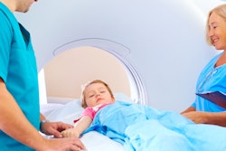
A: In principle, parents can be present during the examinations, although this is more the case for bedside examinations. The MRI examination is usually accompanied by the neonatology staff, who usually also communicate the findings to the parents. Close cooperation between the pediatric radiologists and the pediatric intensive care physicians is therefore particularly important, also in the run-up to the examination. Here, the focus is on joint education of the parents in order to convey to them the necessity of the examination and to take away their fear of it.
Editor's note: This is an edited version of a translation of an article published in German online by the DRG. Translation by Frances Rylands-Monk. To read the original version, go to the DRG website.


