On an evening in early September 1971, two men met at a fast-food restaurant for a hamburger dinner in the small town of New Kensington in Pennsylvania. One of them was Paul C. Lauterbur, a professor of chemistry in charge of the nuclear magnetic resonance (NMR) laboratory at the State University of New York at Stony Brook. The other man was Don Vickers, another NMR scientist.
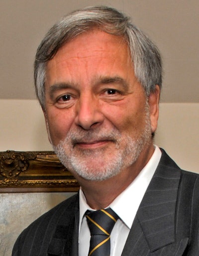 Dr. Peter Rinck, PhD.
During the dinner, Lauterbur explained to Vickers his idea to create images with NMR equipment -- an idea he further developed during the meal. The concept sounded simple in theory: Superimpose on the strong magnetic field of an NMR spectrometer a second smaller and adjustable field.
On the next day, Lauterbur bought a laboratory notebook and put down in writing the background and outline of "Spatially resolved nuclear magnetic resonance experiments," signed the text, and had it witnessed by Vickers on 3 September 1971.
Early development of NMR
Magnetic resonance -- or NMR as natural scientists call it -- is a phenomenon that was first mentioned in the scientific literature before World War II. In 1946, independently of each other, two scientists in the United States described a physico-chemical phenomenon that was based upon the magnetic properties of certain nuclei in the periodic system. The two scientists, Edward M. Purcell and Felix Bloch, were awarded the Nobel Prize in Physics in 1952.
Purcell and Bloch found that when these nuclei were placed in a magnetic field, they absorbed energy in the radiofrequency range and reemitted this energy during the transition to their original orientation. Because the strength of the magnetic field and the radiofrequency must match each other, the phenomenon was called NMR: nuclear because it is only the nuclei of the atoms that react, magnetic because it happens in a magnetic field, and resonance because of the direct dependence of field strength and frequency.
Before Lauterbur's discovery, nobody could determine from where within a sample the NMR signal stems. It could originate at the left or right end, at the top, or at the bottom. Lauterbur's new technique changed this. He joined the strong magnetic field and a second weaker field, the gradient field.
Because the strength of the magnetic field is proportional to the radiofrequency, the frequency of, for instance, a hydrogen nucleus of a water molecule at one end of a sample differs from the signal of another hydrogen nucleus at the other end of the sample. Thus, the location of these nuclei can be calculated. Once their location is known, an image can be created of a slice through an object or in three dimensions of the entire object.
Although Lauterbur did not suggest distinct applications of the new technique in his paper, he did refer to the fact that it had been shown that some "normal" tissues had different signal properties compared with pathological tissue, and he believed that his technique could be used for medical imaging. Thus, he urged his university to file a patent application, but because neither the university patent lawyer nor the university administration itself believed in his idea, no patent application was filed and Lauterbur never obtained a patent on his invention.
In the earlier years, several people had described possible applications of NMR in medicine and biology. Erik Odeblad was the first of them. In 1953 he had met Felix Bloch in Stanford. Odeblad asked him whether he could use his NMR spectrometer to study human samples, but Bloch's response was negative. He made it clear that NMR was a tool for physicists, not for research into physiology, medicine, or biology. Odeblad returned to Sweden and got his own machine.
The two most important scientists for the development of magnetic resonance in medicine and biology were Erik Odeblad, who in the early 1950s first described the differences of relaxation times in human tissue,1 and Paul C. Lauterbur.
Latuerbur faces rejection
Lauterbur stumbled when he tried to publish his invention. In late 1972 he received an apologetic letter from the editor of Nature that read as follows:
"With regret I am returning your manuscript which we feel is not of sufficiently wide significance for inclusion in Nature. This action should not in any way be regarded as an adverse criticism of your work, nor even an indication of editorial policies on studies in this field. A choice must inevitably be made from the many contributions received; it is not even possible to accommodate all those manuscripts which are recommended for publication by the referees."
The paper submitted was very short and described his new imaging technique he had dubbed zeugmatography. For those who did not study Greek at school, zeugma is the yoke, or as the author put it: "That is what is used for joining."
Lauterbur replied: "Several of my colleagues have suggested that the style of the manuscript was too dry and spare, and that the more exuberant prose style of the grant application would have been more appropriate. If you should agree, after reconsideration, that the substance meets your standards, ... I would be willing to incorporate some of the material below in a revised manuscript ..."
The answer from the editor was short and positive: "Would it be possible to modify the manuscript so as to make the applications more clear?"
Finally, the paper was accepted and published in the 16 March 1973 issue of Nature under the title "Image Formation by Induced Local Interaction: Examples Employing Magnetic Resonance."2
The Nobel Prize
Thirty-two years after his invention, in 2003, the Nobel Committee conferred their Prize in Medicine on Lauterbur for the invention of magnetic resonance imaging. He shared it with Peter Mansfield, a British physicist, who was recognized for the further development of the technique. This was the first Nobel Prize in Physiology or Medicine awarded in the field.
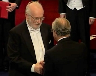 Lauterbur receives the Nobel Prize from the King of Sweden. Copyright: The Round Table Foundation.
Lauterbur commented on this in a lecture given in Lund, Sweden, some days after the Nobel Prize Ceremony in Stockholm in 2003:
"It has been noted that the Nobel Prize for the development of MRI was awarded to a chemist and a physicist. That is not accidental. The field developed from a discipline that was first the province of physicists, two of whom shared a Nobel Prize for it, and then became most prominent in its applications to chemistry, so that chemists received the next two Nobel Prizes, for novel techniques and applications. Although the needs of medical diagnosis stimulated the development of MRI, it was firmly grounded in the knowledge and instruments of physicists and chemists, as well as those of mathematicians and engineers, all far from the knowledge and concerns of physicians, who became its greatest beneficiaries."
A short history of MRI can be downloaded free of charge from the Round Table website.
References
- Odeblad E, Lindström G. Some preliminary observations on the proton magnetic resonance in biological samples. Acta Radiol. 1955;43:469-476.
- Lauterbur PC. Image formation by induced local interactions: examples of employing nuclear magnetic resonance. Nature. 1973;242:190-191.
Parts of this article were published 30 years ago as the first Rinckside column, "How it all began." (Rinckside. 1990;1,1:1-3.)
Dr. Peter Rinck, PhD, is a professor of radiology and magnetic resonance and has a doctorate in medical history. He is the president of the Council of The Round Table Foundation (TRTF) and the chairman of the board of the Pro Academia Prize.
The comments and observations expressed herein do not necessarily reflect the opinions of AuntMinnieEurope.com, nor should they be construed as an endorsement or admonishment of any particular vendor, analyst, industry consultant, or consulting group.


