A new view called flexed elbow valgus external rotation (FEVER) improves MRI's ability to evaluate the ulnar collateral ligament in Major League Baseball (MLB) pitchers, according to a study published June 4 in the American Journal of Roentgenology.
Pitchers are vulnerable to ulnar collateral ligament (UCL) injuries due to repetitive throwing motions, a team led by Dr. Pamela Lund of SimonMed Imaging in Las Vegas wrote. But standard MRI positioning for the elbow is often suboptimal for visualizing the UCL.
Lund and colleagues -- including Arizona Diamondbacks team physician Dr. Gary Waslewski -- evaluated the effect that using FEVER view on MRI scans would have on ulnotrochlear joint space measurement.
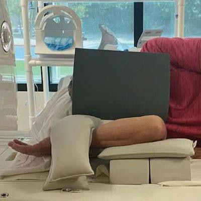
 Note elevated flexed elbow and sandbags to induce valgus stress. Elbow coil is not included in image. Image courtesy of the American Roentgen Ray Society.
The study included 44 MLB pitchers who underwent standard elbow MRI and a FEVER sequence. The researchers found that the FEVER view protocol increased ulnotrochlear joint space width and confidence in three of five UCL-related findings.
"The [results] support the FEVER view as a practical addition to standard elbow MRI protocols for achieving elbow valgus stress in throwing athletes, thereby providing functional information to complement the high-resolution anatomic assessment provided by MRI," Lund and colleagues concluded.


