Using MRI, Israeli researchers have established a set of benchmark measurements for fetal facial and cranial structures to help chart development over multiple gestation weeks. Their findings were published on 12 March in the European Journal of Radiology.
By using eight parameters, including maximal nasal length, the distance between the centers of the eyes, and the space from the forehead to the upper lip, clinicians could better identify potential abnormalities in a developing fetus prior to birth.
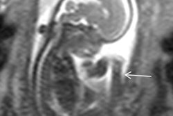
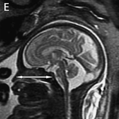
 MR images shows fetal measurements for maximal nasal length between the nasal tip and the posterior wall of the nasopharynx (E, white arrow), mandibular vertebral length (F, white arrow), septal height (G, white arrow), and septal length (H, white arrow). Images courtesy of EJR.
"Novel facial biometric parameters that correlate with [gestational age] hold cardinal information for the prenatal evaluation of facial development and thus surface the need for additional research in order to asses these findings as radiologic markers for facial structural pathologies," wrote Dr. Arik Toren from Sheba Medical Center in Ramat Gan, Israel, and colleagues.
Only a handful of MRI studies have explored the timely progression of fetal facial structures. While the modality can accurately identify and measure many of these "anatomical landmarks," previous studies were "based on a relatively small sample size, and no information is available regarding the nasal cavity and septum," Toren and colleagues wrote. Thus, the team sought to establish a set of standard benchmarks to better assess facial and cranial progression based on gestational age and gender.
The retrospective study included 1.5-tesla MRI scans (Optima, GE Healthcare) from 255 women whose fetuses' brains were imaged from the 24th to 36th week of gestation. Participants were chosen because the fetus appeared normal on first-trimester ultrasound, had a normal facial morphology for gestational age on MRI, and had no intra- and/or extracranial abnormalities. The women were also undergoing MRI scans due to the suspicion of fetal infection, a cerebral abnormality on ultrasound, and reduced fetal movement.
The researchers targeted eight facial features that are considered critical in the normal development of the fetal face and can be easily measured on prenatal MRI. They also can be correlated with gestational age and gender. In this study, the researchers confirmed 134 male and 88 female fetuses.
Toren and colleagues discovered that seven of the parameters correlated significantly with gestational age. The lone exception was the midsagittal view of the fetal profile, known as the inferior facial angle, which runs from the forehead down to the nasal bones and the upper lip. The correlations between these facial parameters and gestational age can be valuable in "temporal prenatal follow-ups to assess facial structure development," they added.
As for gender differences, males had a longer septal length, or the distance between the bridge of the nose at the nasal base and the sphenoid; biparietal diameter, which is the distance between the two temples; and maximal nasal length, which is the distance between the nasal tip and the posterior wall of the nasopharynx. Females, meanwhile, had a wider inferior facial angle.
Additional research with a larger sample size is warranted to further explore the normal development of the cranium and face in fetuses, the authors noted; however, these findings "can provide a reference for prenatal surveillance and influence important decision-making."


