Researchers from the U.K. have developed a new 3D MRI technique for calculating heart muscle strain. They discuss how their method may help improve the accuracy of quantifying cardiac health without the need for gadolinium contrast in an article published online 28 August in Scientific Reports.
Clinicians frequently rely upon an assessment of the myocardium when offering prognosis and ideal management plans to patients with cardiovascular disease. A common way to determine heart muscle health is through myocardial tracking and strain calculation -- two processes that often require patients to undergo a gadolinium-enhanced cardiac MRI protocol, noted lead author Jayendra Bhalodiya, a doctoral candidate from Warwick Manufacturing Group (WMG) of the University of Warwick, and colleagues.
Seeking to quantify heart muscle health without the need for gadolinium contrast, Bhalodiya and colleagues developed a new image-registration algorithm that uses computational mathematics to calculate heart muscle tracking and strain on 3D heart models based on contrast-free MRI scans. They tested the accuracy of their technique on 3D MRI models drawn from 15 separate, publicly available patient imaging datasets.
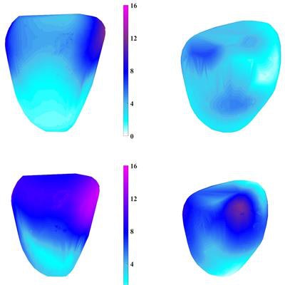
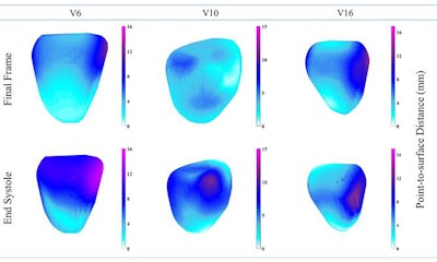 Comparison of heart muscle displacement in the left ventricles of three patients (V6, V10, V16) as calculated on 3D MRI. Image courtesy of the WMG at the University of Warwick.
Overall, the researchers found that the median heart muscle tracking error of their 3D MRI technique was 1.49 mm, which falls within the standard range of errors reported in the literature. In terms of maximum tracking error, the new technique proved to be even more accurate than standard techniques -- averaging a maximum error that was roughly 24% lower than the average maximum tracking error reported in the literature.
"Using 3D MRI computing technique we can see in more depth what is happening to the heart, more precisely to ... heart muscles and diagnose any issues such as remodeling of heart that causes heart failure," co-author Mark Williams, PhD, said in a statement from the university. "The new method avoids the risk of damaging the kidney opposite to what traditional methods do by using gadolinium."
Patients uncomfortable with staying very still for long periods of time in the enclosed environment for gadolinium-enhanced MRI exams may benefit greatly from the 3D MRI approach, Bhalodiya added.
"This new MRI technique also takes away stress from the patient ... [and] the technique doesn't require a dosage of anything, as it tracks the heart naturally."
Though not yet demonstrated in real clinical applications, the 3D MRI technique has the potential to aid in diagnosing heart attacks and recommending certain treatments, such as cardiac resynchronization therapy for patients with a heart valve disease, according to the authors.


