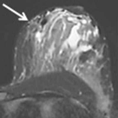
A woman presents with pregnancy-associated breast cancer: How do you image her? Despite high background parenchymal enhancement, MRI is appropriate and accurately spots satellite lesions, according to researchers from Tübingen, Germany. Breast MRI expert Dr. Christiane Kuhl is convinced the study has merit.
A team from University Hospital Tübingen searched the electronic database at the facility to identify women with pregnancy-associated breast cancer and existing breast MRI. Dr. Jana Taron, from the department of diagnostic and interventional radiology, and colleagues evaluated the MR images and compared them with ultrasound and mammography reports. They found breast MRI detects carcinomas and identifies further lesions in the lactating breast.
"By using additional postprocessing methods, i.e., supplemental subtraction images, the high background enhancement can be eliminated to facilitate diagnostics for better tumor visualization," they wrote (European Radiology, 18 September 2018). "Ultrasound remains a reliable and readily available diagnostic tool, while interpretations of mammography can be difficult in these patients due to high parenchymal density."
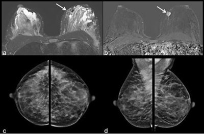
 Images of a 40-year-old patient with invasive ductal carcinoma in the left breast at 9 o'clock. The tumor (white arrow) was displayed in the MR images (a = T2 short-tau inversion recovery, b = contrast-enhanced subtraction image). Due to high density of the lactating breast tissue the mass was occult on mammography (c = craniocaudal and d = mediolateral oblique view of both sides). Windowing of MR images was adapted accordingly. Images courtesy of European Radiology.
Pregnancy-associated breast cancer
Pregnancy hormones cause ductal and lobular growth, leading to an increase in breast size and higher density of glandular tissue. Although about 70% to 80% of all findings detected during lactation are benign, it is of utmost importance to rule out suspicious or malignant masses in the breast as it affects both mother and child, according to Taron and colleagues.
Pregnancy-associated breast cancer is a disease with onset during gestation or within the first year postpartum and accounts for about 3% of all breast cancers or 0.3 in 1,000 pregnancies.
"While it is still very rare, the numbers have been increasing in the past few years, which can mainly be attributed to the increasing age of childbearing women who already present a higher risk for breast malignancies," they noted.
Ultrasound is considered the gold standard -- safe and quick -- while mammography is used more rarely. MRI during pregnancy is problematic but not so postpartum. However, pregnancy changes lactating tissue and enhances parenchyma. Can those difficulties be overcome? That's precisely what the researchers sought to determine.
They investigated the detectability of breast cancer in lactating glandular tissue with a focus on enhancement kinetics of background parenchyma and tumor lesions by using pre- and postcontrast acquisitions and their derived postprocessed subtraction images. They evaluated the detectability of lesions in the postprocessed images and assessed the value of additionally calculated supplemental subtraction images. All of this was compared with ultrasound and mammography findings.
The study included 19 symptomatic patients (ages 27-42) all imaged between December 2005 and December 2017 on a 1.5-tesla MR system (Achieva, Philips Healthcare). The imaging protocol consisted of an axial T2-weighted short-tau inversion recovery (STIR) sequence, as well as an axial dynamic T1-weighted gradient-recalled echo sequence without fat saturation with intravenous application of 0.1 mmol/kg to 0.16 mmol/kg gadolinium chelate (Magnevist 0.5 mmol/mL or Gadovist 1.0 mmol/mL, Bayer Healthcare).
MR images were obtained at the 19th week of pregnancy in one patient, during breastfeeding in four, and after weaning (two days to four weeks) in 14. Time between giving birth and MRI was four days to five months.
| Parenchymal enhancement in lactating women | ||||
| Minimal | Mild | Moderate | Marked | |
| Background parenchymal enhancement | 1 | 3 | 7 | 8 |
Kinetics measured plateau (n = 8), continuous (n = 10), and not quantifiable (n = 1). Tumor kinetics presented washout (n = 17) and plateau (n = 2). Eighteen of 19 tumors were identified on the supplemental subtraction images.
| Visibility of tumors in lactating women | |||
| Ultrasound | Mammography | MRI | |
| No. of tumors visible | 19 (100%) | 12/19 (63.2%) | 19/19 (100%) |
MRI identified additional lesions in six patients, two of whom were reassessed due to therapeutic relevance by second-look ultrasound and characterized as malignant.
"In this evaluation, MRI of the lactating breast was proven to be a reliable diagnostic tool in the assessment of suspected breast neoplasms," Taron and colleagues wrote. "Our study includes 19 patients with pregnancy-associated breast cancer, all of which were detected in the MR images regardless of the amount of background enhancement."
Regarding the use of supplemental subtraction, they identified malignant lesions in 18 out of 19 patients, leading to a detection rate of 95%. When comparing conventional versus supplemental subtraction images, the researchers found no difference in measured tumor size.
"No further lesion was detected in the additionally calculated supplemental subtraction images in our collective," they wrote. "Nevertheless, this method of reducing the background parenchymal enhancement to a minimum and displaying the area of washout kinetics can be of great help to securely detect a malignant lesion, rule out satellite lesions, or identify tumor spread in the surrounding tissue, especially when background parenchymal enhancement is marked (which is the case in lactating women)."
They consider supplemental subtraction a "supporting tool" to ensure diagnosis.
 Dr. Christiane Kuhl.
The current study has some limitations, particularly the small number of patients. Also, final correlation of tumor size measured in imaging and confirmed by histology was only possible in a minority of patients. Most patients received neoadjuvant chemotherapy to reduce tumor size before final surgery.
Nonetheless, the study has merit, according to breast MRI expert Dr. Christiane Kuhl, the clinical director for diagnostic and interventional radiology at Aachen University Hospital in Germany.
"What this study adds is that lactation-induced changes appear rapidly reversible after weaning. And this is of course encouraging," she told AuntMinnieEurope.com. It's encouraging primarily because so far, breast MRI is discouraged in women who are lactating. This study demonstrates that need not be the case.


