_20180817182650.png?auto=format%2Ccompress&q=70&w=400)
Cancer chemotherapy is well known to cause toxic side effects and so, unsurprisingly, scientists are keen to develop a targeted delivery system that avoids adverse impact on healthy tissue. One attractive targeting mechanism is to magnetically guide nanocarriers of toxic drugs to tumors using an externally applied magnetic field. Magnetic targeting is cheap, easy to handle, and versatile; however, it currently lacks a quantifiable method for evaluating targeting efficiency within patients.
MRI is a noninvasive method for in vivo detection, producing 3D images proven to enable quantification of contrast agents such as gadolinium, for example. Now a French research collaboration has proposed a new method to process MR images for iron oxide quantification.
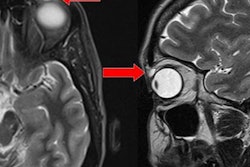
_20180817182703.png?auto=format%2Ccompress&fit=max&q=70&w=400) In vivo MR images of tumors before (a) and after (b) injection of ultramagnetic liposomes, with and without magnetic targeting. Image courtesy of C J Thébault et al, Mol. Imaging Biol. 10.1007/s11307-018-1238-3.
In vivo MR images of tumors before (a) and after (b) injection of ultramagnetic liposomes, with and without magnetic targeting. Image courtesy of C J Thébault et al, Mol. Imaging Biol. 10.1007/s11307-018-1238-3.The researchers created ultramagnetic nanocarriers and applied magnets to target the nanocarriers to colon tumors in mice. They then developed a novel quantification method based on the modification of intensity distributions found in MRI (Molecular Imaging and Biology, 25 June 2018).
A magnetic target
Liposomes are popular nanocarriers because of their biocompatibility and versatility, but for efficient magnetic targeting, liposomes have to contain high levels of iron oxide. Researchers in the group of Christine Ménager, from Laboratoire Phenix in Paris, had previously achieved high loading of magnetic nanoparticles of maghemite (γ-Fe2O3) into liposomes using the reverse phase evaporation process. Now members of Ménager and Bich-Thuy Doan's teams manufactured these ultramagnetic liposomes for magnetic targeting.
Iron oxide nanoparticles are good T2-weighted MRI contrast agents. Thus the researchers used dynamic susceptibility contrast MRI with a T2-timed schedule of radiofrequency pulse sequences to monitor ultramagnetic liposomes distribution in mice. Initial injections of ultramagnetic liposomes were used to assess the nanocarrier stability and survival time in circulation. Hepatic monitoring showed that total uptake of ultramagnetic liposomes (and therefore its exit from circulation) occurred one hour after injection, and, from this, the ultramagnetic liposomes time in circulation for efficient magnetic targeting was estimated at 30 minutes.
With experimental parameters now established, the scientists implanted CT26 murine colon carcinoma cells into the posterior flanks of Balb/C female mice. After two weeks of tumor growth, mice were put under anesthetic and preinjection imaging acquired.
The researchers then placed magnets over the skin of one tumor, leaving the tumor on the opposite flank of each mouse as a control. They intravenously injected ultramagnetic liposomes, then after 30 minutes the magnets were removed, postinjection MR images taken and the animals sacrificed for ex vivo processing.
Ultramagnetic liposomes signal was observed in nonmagnetic targeted tumors due to passive accumulation. To compare the two types of accumulation, the scientists calculated the pixel intensity in each tumor's MRI slices. Intensities were compiled to give a single pixel intensity distribution per tumor, but comparing the mean signals between tumors didn't show any differences.
Adjusting for intensity
The researchers noticed that ultramagnetic liposome presence in the postinjection images caused a shift in the intensity distribution toward the lower end, and realized this was affecting their results. To adjust for this low-intensity shift and enable standardized comparison between time points and tumors, they calculated the percentage of pixels under the I0.25 value (0.25*(maximum intensity -- minimum intensity) for each tumor in an animal.
This new semiquantitative method was able to distinguish differences between differently treated mice and their tumors. The average I0.25 for tumors that only passively accumulated ultramagnetic liposomes increased from 2.9% (in a reference tumor) to 15.3% in injected mice. Magnetic targeting, however, accumulated significantly more ultramagnetic liposomes in tumors, with 28.6% under the I0.25.
Ex vivo processing of murine tumors and organs validated the in vivo MRI quantification technique. Confocal imaging of a fluorescent tag added to ultramagnetic liposomes lipids confirmed that magnetically targeted tumors had higher ultramagnetic liposomes presence than those without. Iron titration experiments confirmed the same result, showing a three-fold accumulation of iron in tumors with magnetic targeting.
This new method enables swift and robust analysis of nonhomogenous MRI signal on any MRI system. And now they have a tool to quantify targeting efficiency in vivo, the researchers seem keen to trial drug encapsulation within the ultramagnetic liposomes.
The researchers also want to evaluate the accumulation efficiency in different tumor types and suggest their method could be applied to compare other targeting methods that utilize T2 contrast nanocarriers.


