Contrast-enhanced ultrasound (CEUS) has many advantages: It's versatile, well-tolerated by patients, and cost-effective, according to Dr. Thomas Fischer, chair of the ultrasound working group (AGUS) of the German Radiological Society (DRG, Deutsche Röntgengesellschaft) and head of the Interdisciplinary Ultrasound Centre at the Charité Hospital in Berlin. In this interview, he explains the main indications for the technology and how CEUS certification can help increase user acceptance.
Why is contrast-enhanced ultrasound so important for radiologists?
Fischer: The growing role of ultrasound within radiology is closely associated with the development of modern sonographic techniques. One of these, CEUS, is now an essential part of our everyday lives. It gives our patients one of the lowest-risk contrast examinations available, with adverse effects in less than 0.02% of cases. Its diagnostic accuracy for space-occupying lesions of the liver and kidneys is at least comparable with a latest-generation CT scan.
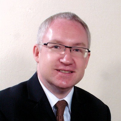
 Dr. Thomas Fischer.
For these reasons, the ultrasound working group wants to increase the role of CEUS in radiology so there's a level playing field with other methods when it comes to making the best possible diagnostic decisions for patients. If you choose ultrasound in a specific situation, you're avoiding radiation exposure and getting a very convincing diagnosis.
Of course MRI is the gold standard in liver diagnostics, for example with the option of a liver-specific contrast medium, and performs slightly better in major comparative studies of diagnostic accuracy. But it's also much more expensive than CT or ultrasound.
If you narrow down the field of application to cases involving a large number of primary benign renal lesions such as focal fatty change, hemangioma, focal nodular hyperplasia, and cysts, major cost-benefit case studies favor CEUS technology even more. But it also takes technical and clinical expertise to use it and achieve a similar quality to other methods.
So far, though, CEUS is not a compulsory part of the radiology continuing education curriculum. Modern ultrasound is the world's most common imaging technique, and there have been a lot of new technical developments so the learning curve is particularly steep. AGUS is holding lectures and practical workshops to address this, and the AGUS board has set up a CEUS certification scheme based on preliminary work by Prof. Teichgräber of Jena.
What are the specific indications for CEUS?
CEUS technology has been evaluated in large diagnostic studies of focal lesions of the liver, and it's proven to be as effective as cross-sectional CT and MRI. Of course, that's only true if the examination of the patient can be easily carried out. So, for example, CT and MRI are better than ultrasound when you're dealing with a massive liver steatosis. It's easy to establish whether this is the case by carrying out a B-scan ultrasound immediately beforehand, and this affects only a small proportion of patients.
As well as this well-known indication, follow-ups after endovascular aortic repair (EVAR) treatment of abdominal aortic aneurysm are playing an increasing part in detecting endoleaks. The advantage is you do a single CT scan after the intervention to assess the condition. You can assess with greater sensitivity and fewer side effects whether the aneurysm sac is shrinking or whether there's an endoleak by using an ultrasound contrast medium, ideally with image fusion technology, which combines the good morphological overview provided by CT and the high spatial resolution of ultrasound.
CEUS can also be used to assess renal cysts just as reliably as with CT. Other new indications include ruling out organ lesions in young, hemodynamically stable patients, and drainage control, for which only three drops of the substance are sufficient to check catheter positioning.
What are the advantages of CEUS compared with other procedures?
They include extremely good tolerance, lack of radiation exposure, and suitability for patients with renal failure or contraindication for MRI. Comparative studies also show that CEUS has a financial advantage: It is simply more cost-effective than CT or MRI for suitable patients. As hospitals come under increasing financial pressure, a problem that is not to be underestimated, radiologists should have access to this method. CEUS is also a safe alternative when you want to avoid repeat examinations, or iodine contrast media are contraindicated, or you can't carry out an MRI because the patient has a pacemaker.
Another big new application is pediatric radiology. It avoids the radiation exposure of CT scans -- radiation damage is much worse in children than in adults -- and you don't have to sedate the patient, whereas you often do with MRI.
However, the contrast medium isn't authorized in Europe yet, like many other products in pediatrics and pediatric radiology, so we're talking about off-label use. The only exception is radiological diagnosis of reflux in children, where voiding cystourethrography has been replaced by contrast ultrasound. Apart from this application, it can already be used in the U.S. to characterize space-occupying renal lesions in children so the European authorities still have some catching up to do compared with the U.S. Food and Drug Administration (FDA).
Does CEUS also have a role to play in interventional ultrasound?
Definitely. I've already mentioned checking drainage position, but CEUS also reduces exposure times in angiography and can be combined with other techniques, for example to confirm the occlusion of an arteriovenous fistula. There are also complex image fusion interventions, such as biopsying the aggressive area of a prostatic carcinoma, or using CEUS to determine the ablation area in the operating room after successful focal therapy of less aggressive prostatic carcinoma.
The quality of ultrasound scans, including CEUS, depends on the user. How do you approach this challenge?
Basically, all examination procedures are user-dependent, and CEUS is no exception. AGUS set up the CEUS training certification to develop the skills of young radiologists, and to improve competence within further education and training institutions in this specialist ultrasound technique. We've deliberately made the certification process straightforward to show how simple, innovative, and effective this method is. Young radiologists can already get the CEUS certification as part of the diagnostic radiologist training, provided they're members of the DRG.
A good example is training young emergency room colleagues in focused assessment with sonography for trauma (FAST) ultrasound. Here again, a contrast medium is used, so it's fairly easy to check for spleen and kidney damage in hemodynamically stable accident patients. We're holding FAST ultrasound workshops, each lasting a few days, four times a year. These are combined with supervised practical use of the method.
Radiology centers of excellence are also getting CEUS certification. What's happening there?
The training plan involves a two-day tested interactive CEUS workshop run by AGUS, and participants also sit in on at least two CEUS examinations at a center of excellence of their choice. They get the certification when they've provided proof of attendance. We plan to hold at least one workshop a year on the principles and clinical applications of CEUS. To make it as practical as possible, participants will deal with interactive and tablet-based clinical case studies.
The applications used in the workshop are based on the current CEUS guidelines of the European Federation of Societies for Ultrasound in Medicine and Biology (EFSUMB). Apart from the basics of CEUS, they'll also deal with applications, for example in the liver, trauma, interventions, and pediatric cases. We want the certificate to dispel any misconceptions and show that CEUS isn't witchcraft. Once you've carried out the procedure a few times under supervision, it's an important addition to the range of your radiological skills. And ultrasound specialists have a long-term future: They can't easily be replaced by a computer.
Are you planning to expand the certification in future, for example by holding events at the Radiology Congress or involving more centers of excellence?
This year's interactive CEUS workshop will take place in Munich, and we'll be announcing the date on the AGUS website. We're looking at whether the format can be transferred to the Radiology Congress. Basically, AGUS would welcome there being more CEUS centers across the country to be able to provide comprehensive training. Therefore we're pleased that more CEUS placements are being offered in hospital radiology departments and practices. They have to carry out at least a hundred CEUS examinations a year as part of their clinical routine in at least one area of application, and the numbers of examinations have to be formally confirmed by the head physician or center manager.
Editor's note: This is an edited version of a translation of an article published in German online by the DRG. Translation by Syntacta Translation & Interpreting. To read the original article, click here.


