Researchers have proposed a novel approach to chronic wound healing that involves image analysis combined with 3D modeling and bioprinting. They have used wound images from patients with diabetes, burns, and metabolic conditions that can cause tissue death.
Peyman Gholami and collaborators from the Amirkabir University of Technology in Tehran, Iran, have introduced a framework to produce biologically compatible 3D prints of deep chronic wounds. Such custom-made wound fillings could potentially aid with the healing process.
The research group aims to provide a proof-of-concept that integrates various technologies into a unified solution. Such technologies include the almost automatic identification of the wound, the automatic creation of a 3D model, and the bioprinting of this model. While these technologies are established in their respective fields, it is their merging that provides a semiautomatic solution to chronic wound healing.
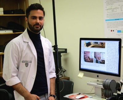 First author Peyman Gholami from Tehran.
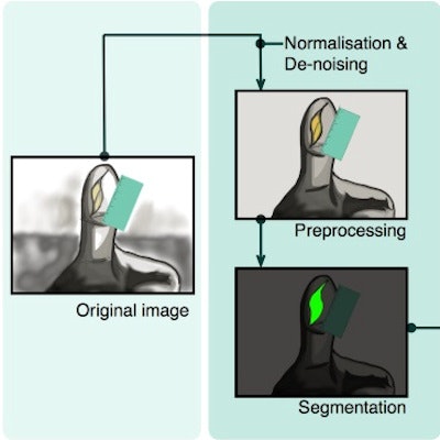
Gholami's work begins with an image of a chronic wound found on the body. After applying the preprocessing procedure, the user segments the wound using the LiveWire algorithm, which creates a mask of the wound with minimal user interaction. Once this mask is generated, the image is calibrated to measurements of the height and width of the wound, matching the pixels in the image to the actual size in millimeters.
Alongside the measurements of the wound, external measurements of the depth are input into a software system that produces a 3D model of the wound that is compatible with a 3D printer (IEEE Journal of Biomedical and Health Informatics, 23 August 2017).
The group chose the LiveWire algorithm following comparison with other common segmentation techniques, due to the high values it achieved on baseline performance indicators, including a score of over 97% in accuracy. The other segmentation algorithms examined included region-growing, active contours, Level set, and texture segmentation.
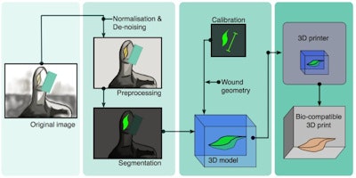
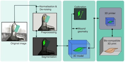
The framework workflow: An image of a chronic wound is computed to obtain a mask of the wound, which is used to generate a 3D model that can be 3D printed.
Using the wound coordinates generated from the segmented image and depth information, the researchers produced the computer instructions, containing G-code, for the bioprinting robot. They printed the wound model using bio-ink hydrogel, which consisted of alginate and gelatine, and was then used for cell encapsulation.
Wounds that won't heal
A recent study on chronic wound detection explains that dealing with chronic wounds is a challenging task for skin pathologists and dermatologists (Wound Repair and Regeneration, Jan.-Feb 2016, Vol. 24:1, pp. 181-188). In normal scenarios, a wound evolves through a set of phases that signify different processes being carried out. Chronic wounds comprise cases where the wound is stuck in one of the phases and doesn't close or heal properly. Causes include factors such as poor circulation, but can sometimes be linked to other conditions.
Gholami and his collaborators used wound images from patients with diabetes, burns, and metabolic conditions that can cause tissue death. The proposed framework provides a good approach for producing custom-made wound fillings.
José Alonso is a doctoral student contributor to medicalphysicsweb, working in the Research Centre for Biomedical Engineering at City, University of London. He is working in biomedical image analysis dealing with immune cell tracking and shape analysis.
© IOP Publishing Limited. Republished with permission from medicalphysicsweb, a community website covering fundamental research and emerging technologies in medical imaging and radiation therapy.
