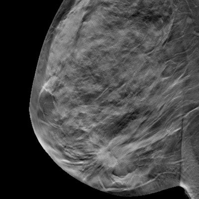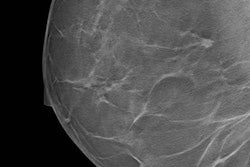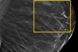
New research from Italy shows digital breast tomosynthesis (DBT) "modestly" increases radiation dose for patients compared with full-field digital mammography (FFDM). However, don't let that be a deterrent in using DBT, the researchers said.
They compared DBT and FFDM radiation dose in more than 1,200 women, finding DBT increases radiation dose by approximately 38%. However, DBT adds clinical value, so radiologists should continue to use the modality, according to a team led by Dr. Gisella Gennaro from the radiology unit at Veneto Institute of Oncology in Padua.
Continue using DBT, especially if acquiring synthetic 2D images, which are 2D images acquired from DBT slices, they noted.
"Assuming an increasing use of synthetic 2D images with DBT (instead of using standard 2D in combination with DBT), and the favorable detection metrics from DBT, the modest increase in radiation dose by tomosynthesis should not constitute an obstacle to its use if there is potential clinical benefit," the authors wrote (European Radiology, 17 August 2017).
Going head to head
DBT continues to increase in popularity and utility. The modality detects more breast cancer and reduces recall rates. Plus, it improves sensitivity and specificity. A major drawback? It increases radiation dose, but by how much? That's precisely what Gennaro and colleagues sought to discover.
They randomly selected a subset of 1,208 patients from the screening study STORM-2, which prospectively compared standard mammography alone, a combination of FFDM with DBT, and a combination of synthetic 2D mammography and DBT. All examinations were performed with the same Selenia Dimensions system (Hologic). Images were acquired in combo mode, which guaranteed the same breast positioning and compression pressure, and it made dose comparisons feasible.
For this study the researchers did not examine the combination of FFDM with DBT, or synthetic 2D mammography plus DBT. They only measured the mean glandular dose for FFDM and DBT.
The researchers included 4,780 FFDM and 4,798 DBT images in the study. They used unprocessed DICOM images to determine mean glandular doses and compared radiation doses between the two modalities by per-view analysis.
The group found statistically significant differences between FFDM and DBT mean glandular doses for all views.
| Dose comparison by view | ||
| Mean glandular dose | FFDM | DBT |
| Craniocaudal | 1.366 mGy | 1.858 mGy |
| Mediolateral oblique | 1.374 mGy | 1.877 mGy |
Bland-Altman analysis showed the average increase of DBT dose compared with FFDM is 38%, with a range between 0% and 75%.
Analyzing mean glandular dose ratios, the researchers found DBT dose increased by between 29% and 40% (median values) for compressed breast thickness within 60 mm, while the increment increased for thicker breasts (45% for 61 mm to 70 mm thickness, 52% for breast thickness > 70 mm). This makes sense because DBT acquisition is performed without an antiscatter grid, and an extra dose is necessary for thicker breasts to compensate for the negative effect of the increased amount of scattered radiation, according to the researchers.
"In general, whatever the dose difference per view, the amount of dose increase due to tomosynthesis is mainly driven by the clinical protocol applied, i.e., it depends on how many DBT views are acquired in addition to standard mammography," they wrote.
For instance, if DBT is used as a "problem solver" in the case of a suspicious mammogram, it might be performed only on the breast where the suspicious finding was detected and restricted to one view. In that case, the total dose increase due to the DBT view would be limited. On the other hand, in screening studies in which both FFDM and DBT were performed, the total radiation dose is at least twice the dose delivered for mammography or even higher.
The current study by Gennaro and colleagues is based on clinical data obtained by a single imaging system, so the generalizability of their research is limited, they noted.
"Further studies will be necessary, with data produced by different FFDM/DBT systems, to have a full picture of dose comparison between tomosynthesis and mammography," the authors concluded.


