The combination of SPECT/CT and fluorescence imaging could help surgeons differentiate tumor tissue from normal tissue in patients with colorectal cancer, according to a paper published in the May issue of the Journal of Nuclear Medicine.
Researchers from the Netherlands used a mouse model to show that pulmonary micrometastases of colorectal cancer can be identified with labetuzumab labeled with both a near-infrared fluorescent dye (IRDye800CW) and indium-111 (JNM, May 2017, Vol. 58:5, pp. 706-710).
Labetuzumab is an antibody developed by Immunomedics that targets carcinoembryonic antigen (CEA), which is overexpressed in approximately 95% of colorectal cancer cases, according to a release from the Society of Nuclear Medicine and Molecular Imaging (SNMMI).
Led by Dr. Marlène Hekman of Radboud University Medical Center, the researchers found that dual use of SPECT and fluorescence after injection of indium-111-DTPA-labetuzumab-IRDye800CW was able to detect the tiny CEA-expressing nodules and could be used to guide resection of tumor lesions.
The approach is applicable to other forms of cancer using a different tumor-targeting antibody, according to Hekman.
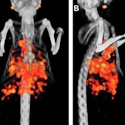
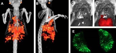
 Example of dual-modality imaging in a mouse one week after tumor cell injection. Pulmonary tumor lesions in the liver and lymph nodes are visualized with micro-SPECT/CT (A, B). After resection of the thoracoabdominal wall, no tumor lesions were visible with the naked eye (C). Fluorescence imaging identified the superficially located tumor lesions (D), and fluorescence imaging of the resected lungs visualized numerous pulmonary lesions (E) (L = lung, H = heart). Images courtesy of Dr. Marlène Hekman and JNM.


