Taking a broader approach to research, combined with much closer collaboration between radiologists and epidemiologists, can contribute to better patient care and disease prevention, according to speakers at a special session about population-based imaging in Germany, held at RSNA 2015 on Monday.
The so-called German National Cohort is a prospective study of 200,000 subjects with an age range of 20 to 69. Only certain regions are included, and people are invited randomly to the study in these areas until the final number has been reached. A total of 18 sites have been established, and more than 300 doctors, nurses, technicians, and assistants are working in these centers.
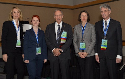 Speakers and moderators during Monday's "Germany Presents" session. From the left are Drs. Sabine Weckbach, Katrin Hegenscheid, Ronald Arenson, Gabriele Krombach, and Norbert Hosten.
"We choose the topic to show how a scientific study can directly influence patient care by radiologists," noted session moderator Dr. Norbert Hosten, immediate past president of the German Radiological Society (DRG) and director and professor at the Institute of Diagnostic Radiology and Neuroradiology at the Medical School of Greifswald University. "Better normal ranges for contrast agents, for volumetrics of liver, heart, etc., are just two examples."
He thinks the scale of the joint effort between radiologists and epidemiologists in Germany is worth close inspection, and is keen to boost radiologists' awareness of the importance of such studies. He urges everybody to encourage their patients to take part in such studies to help improve radiological diagnoses.
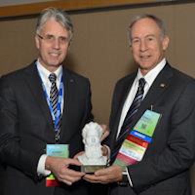
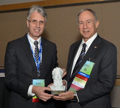 Dr. Norbert Hosten, immediate past president of the German Radiological Society, receives an award from RSNA President Dr. Ronald Arenson.
The central aim of the initiative is to learn about aging and the related changes, according to fellow moderator Dr. Gabriele Krombach, professor and chair of radiology at Giessen University Hospital.
"We are seeking to identify previously unknown risk factors," she told AuntMinnieEurope.com. "And we want to discriminate between the influences of genes and lifestyle. What is the impact of our genes, and what portion is modified and modulated by lifestyle and thus related to 'epigenetic' processes?"
The study will have three levels. All subjects will be involved in the first level, which will take around 2.5 hours for each subject. Subjects are interviewed about almost every aspect of their heath. Cardiovascular exams are performed, blood samples are taken, and cognitive tests are performed. Bone density is also measured.
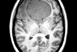
Level two includes even more tests and takes about four hours, but only 40,000 of the 200,000 subjects will be involved. They get an additional exam of their musculoskeletal system, oral exam, ophthalmologic exam, and tests for physical fitness. In level three, 30,000 subjects will receive MRI without contrast medium. Five 3-tesla MRI scanners (all of the same type) have been purchased, and they are located at five of the 18 sites across Germany.
 Certified and well-trained radiologists must be aware of the consequences of reporting incidental findings, according to Dr. Sabine Weckbach.
"Tremendous effort has been undertaken in order to bring them in line regarding similarity of imaging at each of the five sites," Krombach said. "Exactly the same sequences in the same order with the same type of coil will be performed at all sites. The imaging protocol includes T2 and T2-weighted sequences, T1- and T2-mapping of the myocardium, cardiac function, head, lung, and abdominal imaging, and skeletal imaging."
The images are evaluated in the classical way: radiologists read them in order to assess visible signs of diseases, such as tumor formation or enlargement of lymph nodes. Then data are evaluated with software, and imaging biomarkers are evaluated, as is done in radionomics, she continued. Recruitment will take up to five years, and after recruitment has been finalized, all subjects will be reassessed (second round). This will provide knowledge about physiological aging, risk factors, and the course of (degenerative and other) diseases. The hope is to get information that can be used for prevention of diseases.
Dealing with incidental findings
Incidental findings are a big issue here. If a malignant tumor or another serious problem is identified, the subject will be informed about the finding so that he or she can get treated for the disease. This will provide the subject, who now becomes a patient, with the chance for an improved prognosis.
In her RSNA talk, Dr. Sabine Weckbach, a radiologist from Heidelberg University Hospital, made these key points about the role of incidental findings in clinical and research imaging:
Incidental findings in radiology are considered all findings that arise in the context of radiological diagnostics, potentially affecting the health of a subject, without there being an intention to detect the corresponding finding.
- The prevalence of incidental findings is increasing due to the wider usage of techniques such as MRI and CT in routine clinical practice, as well as the inclusion of imaging such as whole-body MRI in large population-based cohorts.
The reporting of radiological incidental findings may lead to further (even invasive) diagnostics and treatment, and potential psychosocial, occupational, and insurance-related consequences.
- The management of incidental findings in clinical routine is regulated by guidelines of the different academic societies. In the research setting, it differs strongly depending on various factors such as study design, age, and health status of enrolled subjects, imaging modality, etc.
The general consensus is that incidental findings must be disclosed to the imaged persons if the radiological incidental findings are potentially clinically relevant. However, subjects must be also protected from the consequences of false-positives findings.
"The best approach to look at incidental findings is to have certified and especially trained radiologists that are aware of all consequences of reporting these findings," she said.
Big data's role
The organizers of the National Cohort hope to be able to detect imaging biomarkers to help determine the risk of developing a disease, but tools and software are essential to manage the data, according to Krombach.
"Big data plays a key role here," she stated. "We will have to statistically evaluate relations between all the different data obtained such as results of blood samples and genetic testing, exams, and interviews with imaging features. It is important that radiology deals with the topic of big data. We cannot let slip this important topic and give it to epidemiologists alone, and we need to collaborate with specialists from informatics and computer sciences to handle it."
Imaging of the heart plays a major role in the project. In addition to T1 and T2 mapping of the myocardium, cardiac function will be measured, and normal values will be derived. These values will be assessed according to age-related factors, and even gender and weight, she added.
 Dr. Gabriele Krombach grew up in Remscheid and visited the Röntgen Museum many times during her childhood and as an adult.
"It is the domain of radiology to interpret and analyze MRI and to do image-based research. For radiology, it is an important opportunity, and we must actively search for answers concerning questions related to physiologic (normal) and pathologic aging," Krombach commented. "What is pathologic aging? You might define it as a result of too many risk factors, or in other words, lifestyle ... There are many questions to answer for radiology, which we usually do not ask in our routine work."
This is a big opportunity for radiology, she believes. Radiologists can bring their experiences to the table to help answer big questions facing humanity. For instance: How can we further identify risk factors for rapid aging? Can we identify risk factors that can be avoided so that the physiological process of aging slows down?
Also, there is an urgent need to identify imaging biomarkers that allow radiologists to assess prognosis of the course of chronic diseases. "Not only will we gain knowledge that will help all of us by helping to identify and further avoid risk factors, but also for an individual patient, such knowledge will help to give individualized instructions for a certain lifestyle to increase the health of that specific person and help the individual to prevent diseases as long as possible. In a sense, that symptom-free time is extended. The study will help to make old age a healthy time of our lives for as many people as possible," she pointed out.
Röntgen's birth house
The opening lecture at the special session, "Röntgen, an x-ray journey," focused on the birth house of Wilhelm Conrad Röntgen, which is located in Lennep, part of the midsized city of Remscheid. The DRG purchased the birth house for a euro last year, and plans to spend 1 million euros on renovating it. So far, it has raised half of this sum. Eventually, the building will serve as a meeting place for national and international radiologists and scientists, and will have meeting rooms and guest rooms.
"The birth house of Wilhelm Conrad Röntgen should be considered the possession of our radiological community, and preservation of this precious building should be a common goal of every radiologist," said Krombach, who grew up in Remscheid and visited the Röntgen Museum many times during her childhood and as an adult.
For further details on how you can help, click here.


