What if you had a lung cancer screening modality that was better than x-ray and nearly as good as CT, but with less radiation and expense? Researchers from Hungary are investigating the idea with a digital tomosynthesis system that they assembled themselves, along with homegrown computer-aided detection (CAD) software.
Two prototype tomosynthesis systems have been developed and are already participating in clinical trials, researchers report in a study published on November 12 in Radiation Protection Dosimetry. One trial is comparing tomosynthesis with CT for lung cancer screening, while the other is designed to follow up patients with advanced lung cancer for large-airway stenosis.
Based on preliminary results in a pilot project, the tomo-CAD combination has produced good results, according to the researchers, although as with most CAD studies, they are finding it a challenge to balance sensitivity against false positives.
The promise of tomosynthesis
For the past decade or so, researchers have held up tomosynthesis for chest applications (as opposed to its close relation, digital breast tomosynthesis) as an exciting technology with the cost and radiation dose of x-ray and sensitivity close to that of CT. Previous research has focused on using tomo as a problem-solving tool for suspicious nodules first seen on radiography, before they are sent on to CT.
But the rise of CT lung cancer screening has generated another question: Could tomosynthesis be the ideal screening tool? It lacks the cost and radiation burden of CT, and it could have higher sensitivity than radiography, which has largely failed to prove effective as a screening tool in population-based clinical trials.
To that end, a group including researchers from Hungarian medical device manufacturer Innomed Medical, as well as the Budapest University of Technology and Economics and Semmelweis University, began investigating tomo-based lung cancer screening using tomo and CAD technologies they developed themselves.
The group first built a titling table, MRX NG II, for the tomo system, which supports a 43 x 43-cm digital detector that can move around the patient, in parallel but opposite directions from the x-ray tube. The system acquires up to 60 projection images in a 40° arc over 10 seconds for a full tomo exam, with a spatial resolution of 1,500 x 1,500 pixels.
In parallel, the group developed a CAD algorithm to use on the tomo images. The researchers noted that while CAD for chest CT is fairly routine, tomo CAD is a relatively novel field. They designed their algorithm for two main functions:
- Finding suspicious nodules in the range of 5 mm to 25 mm in diameter
- Estimating the volume of detected nodules
Developing a CAD algorithm for tomosynthesis presents a number of challenges, as tomo images fall somewhere in appearance between x-ray and CT, the researchers noted. Tomo systems create slices like CT systems do, but slice thicknesses are much larger.
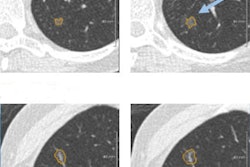
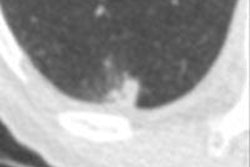
In a series of 27 tomosynthesis scans performed on patients at Semmelweis University, the researchers found sensitivity of 70% to 90% for nodules 3 mm to 7 mm, with one false-positive detection per tomo slice. They concluded that this was far too many false positives, so they are considering the development of new algorithms.
With respect to measuring nodule volume, the researchers noted that this is an important task, as nodule growth over time can help distinguish between aggressive and slow-growing cancers. Their CAD algorithm struggled in measuring lesion doubling time in nodules smaller than 6 mm.
Finally, the group measured the radiation dose delivered by the tomosynthesis unit, since tomo's lower radiation burden is a major selling point of the technology relative to CT. The estimated effective dose of the system for a 60-projection tomo exam was 0.161 mSv, about four times higher than a conventional digital chest exam but only 10% of the dose of a typical chest CT exam.
The researchers noted that there are two studies underway in Hungary with prototype versions of the system. It's being used as a screening tool to detect lung nodules in a trial at the National Koranyi Institute of Tuberculosis and Pulmonology in Budapest, where the goal is to screen 2,000 individuals with tomo and compare its performance to CT. CAD will also be used to analyze images in addition to two independent readers.
In the second trial, being performed at Semmelweis University, researchers are using tomosynthesis in the follow-up of patients with stage III/IV lung cancer who are receiving chemotherapy. Baseline tomo images are being acquired and then compared with scans after treatment to measure changes in the number of lesions, as well as their size and morphology.
Preliminary results from this study indicate that tomosynthesis can be useful for visualizing obstructions in the large airways, which can affect the course of treatment. Tomo can document treatment results and detect restenosis in a way that conventional x-ray can't, the group noted.
"The early results suggest that [tomosynthesis] is a useful method for lung cancer follow-up with the capability of measuring the size and volume of lung nodules and reducing radiation dose," the researchers concluded. "The results of the developed CAD system for finding nodules on the lung fields were also encouraging, although the false-positive rate is still too high."


