An e-learning tool run by radiologists in Switzerland and the U.S. received the only Magna Cum Laude at last week's European Society of Gastrointestinal and Abdominal Radiology (ESGAR) congress. Established in 2010, the Liver Imaging Atlas now has 22,000 users, and 26% of user traffic comes from Europe.
"The atlas is not intended to replace text books on liver imaging or a sound radiological training program," said editor Dr. Orpheus Kolokythas, director of MRI at the Institute for Radiology and Nuclear Medicine, Cantonal Hospital in Winterthur, Switzerland, and affiliate associate professor of radiology at the University of Washington in Seattle, Washington. "The goal is to provide a free, easily accessible, practical, and up-to-date resource for radiologists and other medical professionals in all parts of the world. We are working on an app for tablets and smartphones and it will depend on the funding situation, if we will be able to offer this new service also for free."
','dvPres', 'clsBtn', 'true' );" >
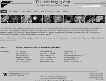
Click image to enlarge.
Unveiled at RSNA 2010 in Chicago, the Liver Imaging Atlas had 4,500 users by early summer 2012, but now has 22,000. This screenshot shows its home page. All images courtesy of Dr. Orpheus Kolokythas and Dr. David Coy, PhD.
Cross-sectional liver imaging, with its various modalities and techniques, is one of the most complex disciplines in abdominal radiology, and a key task is to analyze liver pathology. Each of the three currently leading modalities -- CT, MRI, and ultrasound -- offer specific advantages, but also have their own complexities and limitations, and the large variety of disease entities encountered in the liver, as well as their coexistence, pose a significant challenge to the radiologist, according to Kolokythas and fellow editor Dr. David Coy, PhD, an attending physician and radiologist at the Virginia Mason Medical Center in Seattle.
"Education in diagnostic radiology very much depends on visual memory and instructive images; case-based teaching has been an important and much desired way of teaching in this arena, since it allows the visual memory to apply to authentic cases," they noted in their ESGAR e-poster, which was by far the most viewed exhibit during the meeting in Salzburg, Austria. "Teaching hospitals nowadays often provide an electronic teaching file system replacing the classic hardcopy libraries. Sharing electronic teaching files through the World Wide Web is already a reality in some institutions, allowing a larger community of learners to benefit from this form of electronic learning."
Their atlas uses a structured tagging approach, and has a comprehensive collection of CT, MR, and ultrasound liver cases. It allows radiologists and other medical professionals to find imaging examples of common and uncommon pathologies, display relevant differential diagnoses, test their diagnostic skills, and compile selections of cases for teaching and sharing.
The user can search for imaging cases not only by the diagnosis, but also by imaging features and diagnostic categories. To enable this multitude of search modes, a Web-based editing and entering tool was developed to allow uploading and tagging of clinical cases into a database. An interface was designed for the user to retrieve cases using various search modes and presentation schemes of these cases. The view modes are thumbnail view, case view, and full-resolution view mode.
','dvPres', 'clsBtn', 'true' );" >
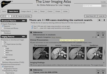
Click image to enlarge.
Screenshot showing an example for 'feature search of all MR cases' that contain the radiographic features 'steatosis' and 'hypervascular' (list of features on the left): 11 cases are being currently returned in thumbnail view mode. The displayed case on top of the displayed case list does not only have CT and MR images, but the patient was also examined over the years with two different MR contrast agents, as indicated on the top right of the case box. All cases are tagged with a diagnosis (in this case: 'adenoma') and with a diagnostic category (here: 'benign neoplasm'), which both function as hyperlinks, so that all cases with that diagnosis or category may be viewed when selected. Tool tips facilitate the use of the atlas. Selecting the view button of each header opens the case in case view mode (see figure below).
A selection of cases or individual cases may be viewed in a training mode (quiz), allowing instructors and trainees to hide the diagnosis temporarily. A bookmark and email feature enables saving and sharing cases for teaching and education. For every search operation in the atlas, one of the three imaging modalities has to be selected. The default search mode when accessing a search page is "CT," until another modality is selected. Case searches can be done by feature, category, index (i.e. diagnosis), and free text search.
In feature search mode, one or more radiographic features may be selected. The features are categorized in structural features, which are shared by the three imaging modalities CT, MR, and ultrasound, as well as imaging features, which may be specific to the modality of choice. The system returns all the cases that are tagged with the selected features. As the search is being narrowed down by selecting more features, all corresponding cases that fulfill the selected criteria will be returned dynamically.
','dvPres', 'clsBtn', 'true' );" >
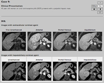
Click image to enlarge.
Screenshot of the quiz function: case view mode of the same case highlighted in previous figure. In case view mode, all images of a case and all modalities can be viewed at the same time, allowing the user to compare imaging features and details across all modalities and contrast phases. In the displayed example, the differences of imaging features between the two different contrast agents on the last contrast phases (equilibrium and hepatobiliary phase) are easily appreciated. In addition, the full text of the clinical presentation and of the imaging findings may be viewed. All images and information may be accessed also in thumbnail view mode as well sequentially, but not simultaneously. In both the thumbnail and case view mode, cases may be viewed in a training mode by selecting the "quiz" button, which temporarily removes all diagnostic clues as shown in this figure: note the diagnosis, diagnostic category, and imaging findings are removed, which allows the compilation of cases for a test environment as well as self assessment. The "DDX" button on the top right opens up a new list of cases with differential diagnoses that share the same features.
The tool was developed at the body imaging section of the department of radiology at the University of Washington Medical Center. It was established in its first version before RSNA 2010.
"The idea was that the radiologist in daily routine, facing a set of images rather than a diagnosis, might be looking for a resource that allows the search for differential diagnoses by imaging criteria, rather than by browsing through possible diagnoses alone," Kolokythas said. "At that time due to limited funding, 'The Liver Imaging Atlas' was more basic with only CT cases available and without the full user functionality that it offers today; however the core feature that cases may be found by either selecting one or more imaging features, or by searching for cases by the diagnosis, or by the diagnostic category has been implemented from the beginning."
In spring 2013, the current functionality level was introduced, with multimodality cases including MRI and ultrasound, and with a substantially expanded set of features including popups with structured information about the various diseases, a quiz mode with various difficulty levels, email, and bookmark functions.
The number of users of the site has steadily increased from 4,500 in early summer 2012 to the current level of 22,000. More than 100 of these are registered users, meaning they can utilize additional free functions such as email and bookmarks.
About 26% of user traffic comes from Europe, 33% from North America (mainly from the U.S.), 30% from Asian countries (largely from India and South Korea); the other usage is evenly spread across the remainder of the Americas, Africa, and Oceania.
Kolokythas and his team are in the process of uploading more cases from Winterthur to the atlas, especially cases with contrast-enhanced ultrasound (CEUS), which is not an approved indication for liver imaging by the U.S. Food and Drug Administration (FDA) at the moment. However, CEUS is an approved, established, and very useful liver imaging technique in Europe and in most other countries around the globe offering unique advantages, warranting its place in the Liver Imaging Atlas along with the other imaging modalities, they emphasized.
Kolokythas went to medical school at the University of Wuerzburg in Germany, and graduated from the University of Heidelberg in 1994. He completed a fellowship in body (abdominal) imaging at the University of Washington in Seattle, and in 2002 became a faculty member and assistant professor at the same institution. He became director of body imaging procedures in 2007 and an associate professor in 2008, and then took up his post as director of MRI in Winterthur in August 2013.
"It is very interesting to observe and experience the differences, but also the similarities in both systems. The Cantonal Hospital in Winterthur is a high-level teaching hospital of the University of Zurich and functions at least at the same academic level as a university hospital," he explained. "There are cultural differences in working life between the U.S. and Switzerland, differences in the reimbursement system, and different disease entities in both continents, which all together enrich the working and life experience of a radiologist working in both worlds."
On his part, Coy is convinced the atlas has great potential as a teaching and learning tool.
"We have tried to leverage the potential of a Web-based platform to maximize the flexibility and customizability of the Liver Atlas to meet the needs of the user -- whether they are a resident learning about liver imaging, a radiologist in practice who wants a quick answer or review, or an educator presenting cases to their trainees -- which is something that cannot be easily achieved by a single book or most other Web-based commercial sites," he said. "By keeping this product free of charge with complete access to anyone with Web access we are able to have users from all over the globe, some of whom may not otherwise have the ability to access commercial resources."
To access the Liver Imaging Atlas, go to www.liveratlas.org.


