Three-dimensional semiautomated software can provide similar measurements and diagnostic accuracy to manual methods for evaluating dynamic myocardial perfusion CT data, a multinational team reported in European Radiology. It's also much faster.
In an article published online on 9 September, a team led by Dr. Ullrich Ebersberger of the Medical University of South Carolina and the Heart Center Munich-Bogenhausen in Germany reported no significant difference in diagnostic accuracy between the two approaches for assessment of myocardial blood flow and myocardial blood volume. In addition, the software took one-third of the time.
"Use of such software substantially decreases postprocessing and interpretation times," the authors wrote. "These results show promise for fostering the integration of this test into clinical workflows."
Labor-intensive approach
While dynamic myocardial perfusion CT has shown promise for adding incremental diagnostic and prognostic information beyond the assessment of coronary artery morphology, postprocessing of this data has been labor-intensive and time-consuming, according to the research team.
"To date, all analyses relied on measurements based on manually prescribed, two-dimensional regions of interest (ROI) within the respective myocardial segment," the authors wrote. "However, such a nonvolume-based approach is vulnerable to substantial variability."
As a result, the group sought to compare the performance and accuracy of a novel 3D semiautomated software package with the standard manual evaluation technique for analyzing myocardial perfusion CT data. Between November 2009 and July 2011, 37 patients were recruited from two centers on two different continents. All patients received pharmacological stress dynamic CT perfusion imaging followed by SPECT within one month.
All CT studies were performed on a second-generation Somatom Definition Flash dual-source CT scanner (Siemens Healthcare). A two-stage protocol consisting of coronary CT angiography at rest and dynamic perfusion imaging during pharmacological stress was performed. Patients received intravenous injection of Ultravist contrast media (Bayer HealthCare Pharmaceuticals).
The dynamic myocardial perfusion studies were reconstructed as transverse sections with 3.0-mm thickness (increment 2.0 mm) and a smooth convolution kernel, according to the authors.
Blinded to the results of the automatic evaluations, two experienced observers then performed traditional manual analysis on Siemens' syngo MultiModality Workplace workstation with syngo VPN perfusion software. The observers, one cardiologist and one radiologist who both had more than five years of experience in cardiac CT, performed their evaluations in consensus.
Image analysis
Prototype software (Cardiac Functional Analysis Protocol Build Data, Siemens) was used to perform semiautomated image analysis. Employing an automatic 3D heart chamber segmentation system and a surface-based four-chamber heart model, the segmentation system combines a flexible heart model with an automatic model fitting approach, according to the researchers.
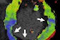
"Using the heart model, an automated chamber segmentation is employed which uses marginal space learning and probabilistic boosting trees for object localization followed by nonrigid deformation estimation," the authors wrote. "The resulting segmentation consequently allowed for both manual placement of ROIs and calculation of a polar map employing the modified 17-segment [American Heart Association] AHA myocardial model."
Manual readjustment of the cardiac chambers can be performed if automatic delineation of the chambers is not done properly.
An E-Cam dual-detector, hybrid SPECT/CT gamma camera (Siemens) was used to acquire SPECT myocardial perfusion imaging studies. Either technetium-99m sestamibi or tetrofosmin were administered intravenously at rest and stress, respectively.
After 19 (3.2%) myocardial segments were excluded due to insufficient coverage and 31 (5.2%) were excluded because of attenuation artifacts, a total of 542 myocardial segments were evaluated in the study. The 3D semiautomated evaluation software successfully processed all CT myocardial perfusion datasets, although additional manual realignment of the endocardial contour was necessary in 20% of the segments.
No difference
The researchers found no difference in mean myocardial blood flow (MBF) and myocardial blood volume (MBV) between the manual approach and the 3D semiautomated evaluation software, as indicated in the table below.
| Manual vs. 3D semiautomated perfusion measurement | |||
| Mean myocardial blood flow | Mean myocardial blood volume | P value | |
| Manual approach | 142.39 ± 47.05 mL/100 mL/min | 142.85 ± 43.23 mL/100 mL/min | 0.5 |
| 3D semiautomated approach | 18.84 ± 5.63 mL/100 mL | 18.60 ± 4.90 mL/100 mL | 0.60 |
Strong agreement was also shown between manual assessment and the 3D evaluation software for both MBF and MBV (Intraclass coefficient [ICC]: 0.91, p < 0.001 for MBF and ICC: 0.88, p < 0.001 for MBV).
The mean evaluation time for MBF and MBV analysis was 49.1 ± 11.2 minutes for manual assessment, compared with 16.5 ± 3.7 minutes for the semiautomated approach. The difference was statistically significant (p < 0.001).
Based on SPECT results, 42 segments in 17 patients were classified as having pathological myocardial perfusion.
| Diagnostic accuracy using manual and 3D semiautomated evaluations | ||||
| Manual approach (MBF) | 3D semiautomated tool (MBF) | Manual approach (MBV) | 3D semiautomated tool (MBV) | |
| Sensitivity | 86% | 79% | 81% | 76% |
| Specificity | 96% | 96% | 96% | 96% |
| Negative predictive value | 99% | 98% | 98% | 98% |
| Positive predictive value | 65% | 65% | 62% | 64% |
| Accuracy | 95% | 95% | 95% | 95% |
"In summary, the performance of 3D semiautomated evaluation software for dynamic CT myocardial perfusion imaging correlates highly with manual assessment of MBF/MBV values in good agreement with SPECT," the authors concluded.


