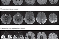
Researchers from Rome have urged all radiologists to boost their knowledge of the MRI findings and characteristics of Creutzfeldt-Jakob disease (CJD), particularly because in some cases MRI can lead to a correct diagnosis even when the clinical information and electroencephalography plus cerebrospinal fluid analysis data are uncertain.
MRI of the brain is the most sensitive technique, and is required in all patients with a clinical suspicion of CJD, noted Dr. Simona Gaudino and colleagues from the Institute of Radiology, "A. Gemelli" Hospital, Catholic University of Rome. In about 80% of cases, CT is generally negative, but it may show rapidly progressive atrophy and ventricular dilatation in 20% of cases.
The DWI (diffusion-weighted imaging) and FLAIR (fluid attenuated inversion recovery) sequences are especially useful, they noted in an e-poster that was awarded a certificate of merit during RSNA 2012 in Chicago. The most common findings are T2 hyperintensity of the basal ganglia, thalamus, and cerebral cortex, while MRI alterations that better correlate with distinct CJD subtypes are DWI/FLAIR hyperintensity of thalamus (variant CJD), pulvinar and hockey-stick sign (variant CJD), and DWI/FLAIR hyperintensity in the occipital cortex (Heidenhain variant).
"CJD is the most common form of transmissible spongiform encephalopathies in humans and occurs worldwide, with an estimated incidence of one case per one million a year," the authors stated. "CJD is reported in almost equal ratios between the sexes, although older males (more than 60 years of age) appear to have a higher incidence of the disease. Previous studies have reported a peak age of onset between 55 and 75 years (mean 61.5 years)."
CJD is a rapidly progressive, fatal, and potentially transmissible disease caused by a prion, which is a proteinaceous infectious particle that lacks nucleic acids, and the active component in prions is an abnormal protein (prion protein, PrP), they explained. Normal animal cells make cellular PrP (PrPC), which is easily soluble and can be digested by proteases. Its expression in the central nervous system is highly regulated during development. Recent studies suggest that this protein has a protective role against dementia and other degenerative problems associated with old age, and it probably prevents death of Purkinje cells and oxidative damage to neurons due to an excess of copper ions.
In animals infected with prions, abnormal PrP (PrPSc) made from normal PrP is highly soluble and resistant to digestion by proteases, and through an autocatalytic process, it promotes the conversion of the cellular PrPc in neighboring neurons, and this leads to further PrPSc accumulation in neurons, according to the authors.
Patients affected with CJD usually present with rapidly progressive dementia, visual abnormalities, and cerebellar dysfunction, including lack of muscle coordination and gait and speech abnormalities. Most patients develop pyramidal and extrapyramidal dysfunction during the course of the disease, with abnormal reflex, spasticity, tremors and rigidity, they continued. Some patients may also show behavioral changes with agitation, depression or confusion. These symptoms often deteriorate very rapidly, and during the terminal stages of illness, these patients develop a state of akinetic mutism. Myoclonus, the most constant physical sign, is present in nearly 90% of CJD cases.
The median illness duration of CJD is four months (mean 7.6 months), and death occurs within 12 months of the onset of the disease in 85-90% of patients, the group pointed out.
New variant CJD, known as "mad cow disease," is a massive common-source epidemic caused by infected meat and bone meal fed primarily to dairy cows with the intention to get high-quality beef. It affects mostly younger people, the median age being 26 years. There are early psychiatric and behavioral symptoms, including limb pain and dysesthesia. As the disease progresses, neurological features become more prominent, particularly ataxia and cognitive impairment. The disease has a median duration of 13 months, they observed.


