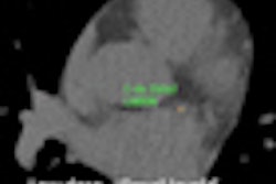
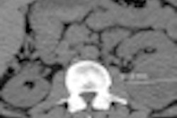
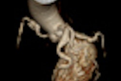
Coronary calcium scoring with an automated technique fits well in a lung cancer screening setting, enabling cardiovascular risk assessment in a population potentially at high risk of cardiac events, according to a Dutch study presented at the RSNA 2011 meeting.
In a study involving nearly 1,800 cases, a team from the University Medical Center Utrecht in the Netherlands found an automated scoring system could be a valuable tool in helping to assign patients to appropriate cardiovascular risk groups.
"Automatic coronary calcium scoring in a lung cancer screening program is feasible," said presenter Ivana Isgum, PhD. In the landmark Nederlands-Leuvens Longkanker Screenings Onderzoek (NELSON) lung cancer detection trial that involved nearly 16,000 participants, more heavy smokers were found to be affected by cardiovascular disease than by lung cancer.
Calcium scoring would seem a good fit with lung cancer screening, which utilizes low-dose, non-ECG-synchronized chest CT scans. Coronary calcium scores from such scans have been shown to be a predictor of cardiac events and mortality, she noted.
Performing manual calcium scoring in a lung cancer screening setting is very difficult, however, due to the large number of acquired images and the CT acquisition protocol; low-dose acquisition produces noise, and cardiac motion can make calcifications either blurry or hard to visualize, she said. The NELSON study also included a population of heavy smokers, with many calcifications per scan that would require a lot of user interaction.
The university built an automatic calcium scoring system, which first creates a coronary calcium scoring map that assigns probability to every location in the scan for the appearance of coronary calcifications. Thresholding is then performed to generate calcification candidates, and a machine learning system completes the process by identifying coronary calcification based on density and location, Isgum said.
To evaluate the performance of the scoring system in a lung cancer screening setting, the researchers selected 1,796 baseline chest CT scans from the NELSON trial.
The study included images from former heavy smokers who ranged in age from 50 to 75, with acquisition parameters of 16 x 0.75-mm collimation and tube current of 30 mAs. The low-dose, noncontrast chest CT images were reconstructed to 3.1-mm sections with 1.4-mm increments.
After automatic scoring was performed, one of four trained observers visually inspected the scans, and, if necessary, provided manual correction. Agatston and calcium volume scores were computed before and after manual correction.
Based on the Agatston score, each subject was then assigned to a cardiovascular risk group. The researchers also wanted to evaluate interobserver agreement for manual scoring, and selected a subset of 45 scans that were manually scored by two observers. This subset was equally divided over the cardiovascular risk groups.
Both methods yielded similar scoring results.
Automated versus manual calcium scoring
|
Automatic coronary calcium scoring more frequently underestimated than overestimated the amount of calcification, Isgum noted.
Based on the volume score, Spearman's rank correlation (ρ) was calculated to assess correlation before and after manual correction of automated scores, and between observers for manual scoring. Linearly weighted kappa statistic (κ) was calculated to evaluate the agreement in cardiovascular risk category assignment.
The researchers found high correlation (ρ = 0.88) and an agreement κ of 0.79 between the automated and manual scores, while interobserver manual scoring had a correlation ρ of 0.89 and an agreement κ of 0.83.
"Agreement between automatic and manually corrected scores is similar to interobserver agreement," Isgum said.


