A computer-aided diagnosis (CAD) system for a physician can be compared to an autopilot: The physician and pilot are both sufficiently educated and experienced to work without the aid of a computer, but a CAD system and autopilot can help them avoid mistakes and support them in difficult situations.
This clear and compelling rationale for CAD is provided by a leading authority on clinical decision support systems, Dr. Lars Edenbrandt, a professor of clinical physiology and nuclear medicine at Gothenburg University in Sweden. He is convinced CAD systems will be of great future clinical value and physicians benefit from the advice of these systems.
CAD applications have so far mainly been used in screening, particularly mammography and CT colonography, and systems have been developed to show a very high sensitivity and to miss very few cases of cancer, he explained. As a consequence, the number of false-positive marks has been very high; about 19 out of 20 marks from a mammography CAD system are false-positive, and the recall rate of patients without cancer is also high.
"We are working with CAD systems in the normal clinical setting, for example bone scans from patients with known cancer and suspected cases of metastases. A more balanced relation between sensitivity and specificity can be used, and, as a result, the number of false classifications by these CAD systems is low," noted Edenbrandt, who also works as a consultant at the Malmö University Hospital in Malmö, Sweden, and is leader of the Lund University Research Programme in Medical Informatics (LUMI).
LUMI aims to coordinate research in the multidisciplinary field of medical informatics. The long-term goal is to develop decision support systems that assist in clinical decision-making. It currently involves about 20 faculty members, up to PhD students, and 20 associate members (engineers, technologists, a research nurse, etc). In 1999, he founded Exini Diagnostics, the CAD software developer formerly called WeAidU. He was CEO of the company until 2007 and is now director of research.
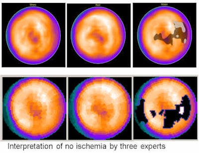
 CAD can assist in the interpretation of myocardial perfusion scintigrams. The top row images were done by Exini, and no ischemia was found. The bottom row images were done by Perfex. The findings were there were four contiguous count deficits in the myocardial perfusion distribution. Count deficits not reported below were deemed to be statistically insignificant at the specified sensitivity and specificity. The second area, which probably is diseased, is mainly located in the lateral wall. There is moderate evidence of reversibility. Source: Dr. Lena Johansson, Dr. Milan Lomsky, Dr. Jens Marving, Dr. Sven-Eric Svensson, Dr. Lars Edenbrandt, P199, EANM congress, 15-19 October 2011.
At Malmö, Edenbrandt and his colleagues have used Exini's bone and heart CAD systems in routine clinical practice for all bone and cardiac scans for several years. The report module of the CAD systems makes it possible to produce a structured report and to export it to the RIS, thereby making it available for referring physicians. CAD has been integrated into the workflow, and reports generated in the CAD systems are directly exported to the RIS. This saves time compared with physicians dictating reports, secretaries typing them up, and physicians proofreading the reports before signing them.
"An interesting experience is that CAD systems are very useful for the normal cases. They help the physician to feel confident that he/she has not missed anything and that there is no need to have a second or third look at the images," he pointed out. "Furthermore, for normal cases the standard report text proposed by the CAD system can be used without changes and the case can be finalized by just clicking 'save.' "
Clinical staff at Sahlgrenska University Hospital in Gothenburg use CAD systems in daily practice as a first or second opinion in complex cases; these patients are normally assessed using Exini heart and other software package. CAD systems are also used as a teaching tool. The results are used by a junior physician during self-teaching sessions, as well as a starting point in a discussion with a senior physician.
Edenbrandt and his colleagues are now developing CAD systems for other types of examinations, including brain and kidney examinations. They are focusing on diagnostic imaging in oncology (bone scans and PET/CT) and cardiology (cardiac scans), partly because these are the most common examinations in nuclear medicine, he noted.
At the recent European Association of Nuclear Medicine (EANM) annual congress, they presented new information about CAD for interpretation of myocardial perfusion scintigrams (MPS). The authors, led by Dr. Lena Johansson from the department of molecular and clinical medicine at Sahlgrenska University Hospital, compared the diagnostic performance of two CAD systems (Exini heart and Perfex) for the detection of ischemia.
They studied 1,052 consecutive patients (499 men and 553 women) who underwent two-day stress/rest Tc-99m sestamibi MPS studies. Patients were stressed using either maximal exercise or pharmacological tests with adenosine. The reference/gold standard classifications for the MPS studies about the presence or absence of ischemia were obtained from three physicians, each of whom had more than 25 years of experience in nuclear cardiology. They re-evaluated all MPS images. The majority rule was applied in all cases of disagreement. No quantitative results from software packages were available during the re-evaluation. Automatic processing was done using the Exini and Perfex software packages.
Ischemia was found in 257 patients by the three experts, and the sensitivity for ischemia was 68.9% for Exini and 62.6% for Perfex. In the remaining patients without ischemia, the specificity was 89.2% versus 82.4%.
"These results show the high potential for neural networks as a computer-aided diagnosis system," the authors wrote. "Exini shows significantly higher sensitivity and specificity for ischemia than Perfex. This difference in performance should be taken into consideration when software packages are used in clinical routine."
To read more about the benefits of using CAD, Edenbrandt has the following suggestions:
Sadik M et al. Improved classifications of planar whole-body bone scans using a computer-assisted diagnosis system: A multicenter, multiple-reader, multiple-case study. J Nucl Med. 2009;50(3):368-375.
Tägil K et al. A decision support system improves the interpretation of myocardial perfusion imaging. Eur J Nucl Med Mol Imaging. 2008;35(9):1602-1607.
Sadik showed in her paper that the 35 physicians involved in the study increased their sensitivity for metastatic disease in bone scans by an average of 10% with the aid of the CAD system, while Tägil showed similar results in a smaller study dealing with cardiac scans, he stated.


