Plain radiography and CT are the most commonly used modalities for imaging of gunshot wounds, but angiography and MRI are playing an increasing role, according to research presented at the 2011 U.K. Radiological Congress (UKRC) in Manchester.
"Penetrating trauma is becoming an increasingly common presentation to emergency departments, especially in larger urban centers," noted lead author Dr. Andrew Shawyer, a radiologist at the St Bartholomew's and Royal London Hospitals, London. "This group of patients has a high mortality and morbidity rate, and accurate diagnosis, injury assessment, and evaluation of complications relies heavily on radiology."
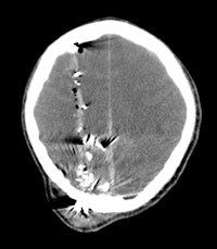
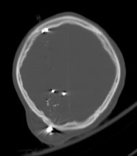 A 30-year-old male with a gunshot wound to the head. Left: Brain window axial CT image demonstrates a significant soft-tissue swelling and underlying fracture at the entry site of the projectile. The course of the bullet is seen by a linear hemorrhage through the right cerebral hemisphere. There is loss of the grey-white differentiation in keeping with a diffuse hypoxic injury. Right: The fragmentation and scatter of the low-velocity projectile is best seen on CT bone windows. All images courtesy of Dr. Andrew Shawyer.
Doctors involved in the care of these patients need to understand the patterns and mechanisms of the injuries, he explained. To accurately interpret images of gunshot wounds, a basic knowledge of ballistics is important, especially the factors affecting the extent and type of tissue damage. Such knowledge is useful not only for evaluating acute injuries but also for determining the path of the missile, awareness of missile fragmentation, and embolization, thus contributing to the overall clinical, and often the forensic, picture.
Gunshot wounds inflicted by handguns are usually classified as low-velocity injuries, having a muzzle velocity less than 600 m/sec, stated Shawyer. In addition to missile velocity, the amount of tissue damage depends on factors such as the design of the projectile (e.g., full metal jacket, soft point, hollow point), biological tissue characteristics, the entrance profile/angle of the projectile when it goes into the body, and the caliber of the projectile.
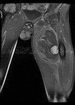
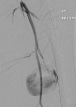
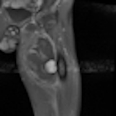
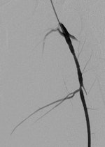 A 19-year-old male with delayed presentation following gunshot injury in the thigh. Left: T1 coronal MRI shows a large pseudoaneurysm in the upper thigh. Pre- (middle) and postembolization (right) angiographic images demonstrate successful treatment of the pseudoaneurysm.
The two mechanisms of tissue injury that account for the majority of damage caused by a bullet are direct crushing of tissue by the projectile (causing a permanent cavity) and temporary cavitation, which stretches and tears surrounding tissues. Increased velocity, fragmentation, deformation, and rolling/spinning of the bullet will cause more damage by both these mechanisms, he pointed out.
Since the 1960s, there has been a steady upward trend in the unlawful use of firearms in the U.K. The use of handguns in criminal acts has grown rapidly since the early 1980s, Shawyer noted. The use of guns in interpersonal violence shows one of the steepest rises, especially between 2000 and 2009, when three police areas -- London, Manchester, and the West Midlands -- accounted for more than half of recorded gun crime in England and Wales. Firearm injuries and fatalities rose from around 2,200 in 1999 to nearly 4,000 in 2002 and more than 5,000 in 2007.
The co-authors of this study were Dr. Wade Gayed, Dr. Bhim Odedra, Dr. Chirag Patel, and Dr. Ian Renfrew.


