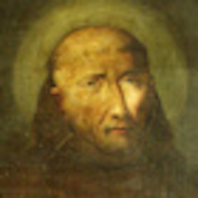
How can the hidden past of artwork be revealed without damaging the creation itself? Since 2003, researchers in Poland have been looking to optical coherence tomography (OCT) for answers, using the technology to detect suspected forgeries in paintings and unearth hidden inscriptions, among other applications.
"Our first research was focused on the examination of paintings, and this has continued to be our main subject. ... The applications for painting are so broad that there is almost no time for other [works]," Piotr Targowski, Ph.D., an associate professor of physics at Nicolaus Copernicus University in Torun, told AuntMinnie.com.
OCT is an optical imaging modality that uses near-infrared light to produce high-resolution cross-sectional images. An optical beam is targeted at an object, and by analyzing the reflected light from multiple scans, researchers can obtain 3D data and a structural view of the object.
Currently being used or evaluated for a variety of medical indications, including vascular imaging, dermatology, oncology, and, in particular, ophthalmology, OCT offers the advantages of being noninvasive and nondestructive -- important features for both medicine and the field of art conservation.
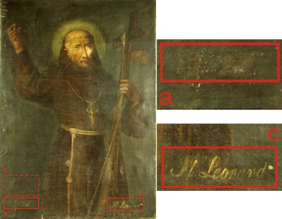 |
| Targowski and colleagues used OCT to clarify the history of the 18th-century painting "Saint Leonard of Porto Maurizio." By analyzing multiple layers of varnish and paint, and where the inscriptions fell among the layers, they concluded that an inscription "1797" (located at area marked a) is likely the creation date of the painting, which also originally carried the inscription "B Leonardus a Maurzio" (located at b). However, after Leonard was canonized, the name (b) was painted over and a new one, "St. Leonard" (c), was added. All images courtesy of Piotr Targowski, Ph.D., and Accounts of Chemical Research. |
"For conservation or inventory purposes, it is very important to reveal the stratigraphy, that is, the order, thickness, composition, and possibly the origin, of the layers building up the painting's structure," which is used for confirming the history of the work, Targowski and colleagues wrote in a recent study (Accounts of Chemical Research, December 31, 2009).
The dilemma, however, is that taking samples from a painting to analyze the layers -- the most common and unequivocal method of stratigraphy -- is by its nature invasive. As a result, the use of sampling is limited by conservation ethics to areas of paintings that are already damaged, which may not give an accurate representation of the work as a whole.
In contrast, noninvasive examination with OCT "may be repeated as many times as necessary, so the results are more representative than from sampling," Targowski explained. Furthermore, "the size of the cross-section is much larger than from sampling -- we usually do scans 7- to 20-mm long, whereas samples are small -- about 1 mm."
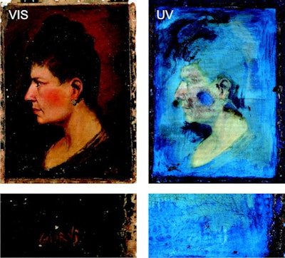 |
| For the 19th-century painting "Portrait of an unknown woman," OCT was used by Targowski and colleagues to investigate a possible forgery. The left images show front illumination of the painting and a closer view of the signature, while the right shows ultraviolet (UV)-excited fluorescence of the same areas. The UV examination indicated that two varnishes are present (fluorescing yellow and blue), and the signature appears to be in an area where the primary varnish was removed before the entire painting was covered by the second (bluish) varnish. Based on OCT results, the group concluded that the original, yellow-fluorescing varnish was likely removed so the signature would appear authentic and placed before any varnish was applied. |
Because of the benefits of noninvasive techniques, other imaging modalities have also been explored for examining paintings, including reflectance spectroscopy in visible light, thermographic techniques such as pulse phase thermography (PPT), air-coupled ultrasound echo detection, and CT, according to Targowski and colleagues.
However, such methods "either are only sensitive to surface properties or do not have sufficient spatial resolution to be useful for structures with an overall thickness of only a fraction of a millimeter: the resulting information comprises an overlap of that from all different depths," they wrote. With OCT, micrometer-level axial resolution can be obtained.
This isn't to say that OCT doesn't have its challenges. The opacity of layers is a primary one -- if a layer strongly absorbs the light, subsequent layers are not visible -- but transparent and semitransparent layers such as varnishes and glazes are easily evaluable. Also, proper interpretation requires a certain level of knowledge and training, Targowski said, and for some objects with very thin layers, OCT's axial resolution still may not be good enough.
Other uses for which OCT has been studied at Nicolaus Copernicus University and by other research groups include analyzing historic jades, ceramics, and glass objects, and evaluating processes such as the drying of varnish layers and alterations in canvas paintings due to environmental changes, according to the researchers.
Although paintings continue to be the main focus for Targowski and colleagues, they also use OCT for examining stained and historic glass, among other applications, and the researchers are interested in the combination of different techniques such as OCT with laser-induced breakdown spectroscopy (LIBS) and laser-induced fluorescence (LIF), he said.
At Nicolaus Copernicus University, the Institute of Physics has cooperated with the Institute for the Study, Restoration, and Conservation of Cultural Heritage on OCT research from the very beginning, Targowski said.
"It is very fortunate that we have the conservation department (and the university museum with a very interesting collection of paintings) at our university," he noted. "Art conservation is indeed one of the specialties of our university -- the only department located in a full-profile university in Poland; other such faculties are located at academies of art. This makes our department more science-oriented."
By Nicole Pettit
AuntMinnie.com staff editor
May 26, 2010
Related Reading
Radiology art reveals beauty -- even in a Big Mac, February 26, 2009
Scan artist: Radiologist uses CT to reveal mystery of antiquities, August 25, 2005
Radiologist at Louvre trades healthcare for high art, November 1, 2002
CT reveals secrets of antique books and collectibles, November 28, 2001
Radiography meets flower power in RT's art, July 17, 2001
Copyright © 2010 AuntMinnie.com


