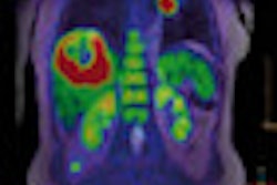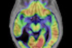
Structural, functional, and molecular imaging of patients with brain tumors is feasible with diagnostic imaging quality using simultaneous hybrid PET/MR image acquisition, according to a study in the August issue of the Journal of Nuclear Medicine.
A pilot study in Germany of 10 patients with intracranial masses found no significant artifacts or distortions using a hybrid scanner configuration that features an MRI-compatible PET insert placed in the magnet bore. The lead author of the study was Andreas Boss, MD, from the department of diagnostic and interventional radiology at Eberhard Karls University Tübingen in Tübingen, Germany (JNM, August 2010, Vol. 51:8, pp. 1198-1205).
As the authors noted, previous studies have indicated that only half of low-grade gliomas can be correctly classified using conventional contrast-enhanced MRI. In addition, conventional MRI may be insensitive to post-therapy changes. With PET's ability to provide biochemical and metabolic information, researchers surmised that the two modalities together "may increase diagnostic accuracy over the single modalities."
Between August 2008 and November 2009, 10 patients with a median age of 51 years (range, 34 to 73 years) participated in the pilot study. All patients initially received a brain PET/CT scan, followed immediately by PET/MRI.
Patient diagnoses
At the time of the patient referrals, diagnoses included three cases of meningioma, two patients with low-grade astrocytoma, two cases of glioblastoma, and one case each of suspected low-grade astrocytoma, anaplastic astrocytoma, and atypical neurocytoma.
For the glial tumors, the radiotracer carbon-11 (C-11) methionine was used for PET imaging. In the cases of meningiomas, gallium-68 DOTA-tyr3-octreotide (Ga-68 DOTATOC) was administered.
Data acquisition from the PET scanner (Hi-Rez Biograph 16, Siemens Healthcare, Erlangen, Germany) started at 30 minutes after the injection of methionine and lasted for eight minutes. When DOTATOC was used, acquisition started at 20 minutes after injection and lasted for four minutes. A noncontrast-enhanced low-dose CT scan was used for attenuation correction in the glioma patients, while a contrast-enhanced CT scan was used for meningioma patients.
PET/MRI exams
All PET/MRI scans were performed with a hybrid PET/MRI system capable of simultaneous PET/MR imaging of the brain and skull base. The system consisted of an MRI-compatible PET scanner (BrainPET, Siemens), which was inserted into a modified 3-tesla whole-body MRI scanner (Magnetom Trio Tim, Siemens).
The researchers acquired PET and MRI data simultaneously, starting 30 to 60 minutes after PET/CT acquisition. The PET scan was conducted for 30 minutes, producing approximately 5 GB of raw data. The PET datasets then were reconstructed by 3D algorithm. Attenuation maps were computed from 3D MRI datasets using an algorithm combining pattern recognition and atlas registration.
Upon review of the resulting images, the researchers determined that the MRI datasets acquired during simultaneous PET acquisition "showed diagnostic image quality, without apparent artifacts or distortions."
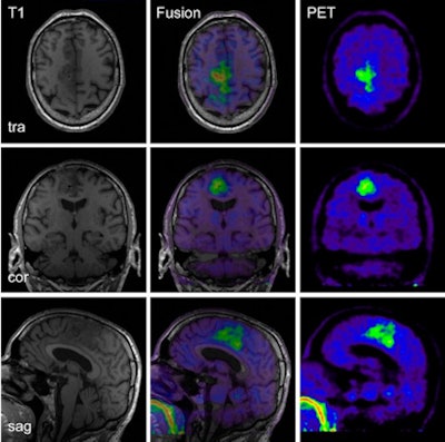 |
| PET/MR images of a 60-year-old patient with anaplastic astrocytoma with right parafalxial tumor extension. MRI and PET datasets were simultaneously acquired at coronal (cor), sagittal (sag), and transverse (tra) sections. Images courtesy of the Journal of Nuclear Medicine. |
The one exception was when a 4-mm Gaussian filter was not used and the PET datasets from concurrent PET/MRI scans "exhibited slight streak artifacts, which were best visible in the sagittal and coronal reformations," the researchers noted. "After filtering, these artifacts were hardly visible."
The group also found that radiotracer decay between the PET/CT and PET/MRI acquisitions was effectively compensated by the longer scanning duration and better scanner sensitivity.
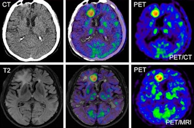 |
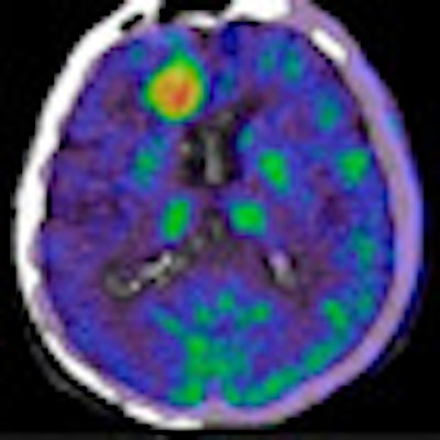
| As an example of similar results with PET/CT and MRI, the images are of a 56-year-old patient with glioblastoma multiforme on the right side in the frontal area close to interhemispheric fissure. Top: PET/CT data show low-dose, noncontrast-enhanced CT scan (left), corresponding fusion image (center), and C-11 methionine PET image (right). Bottom: PET/MRI data show T2-weighted FLAIR image (left), fusion image (center), and PET image (right). |
"Our study indicates that hybrid PET/MRI of intracranial tumors using C-11 methionine or Ga-68 DOTATOC can be reliably performed," Boss and his colleagues wrote. "The image quality and quantitative data achieved using PET/MRI is similar to that using PET/CT."
In addition, the authors noted that the combination of PET with MRI, instead of CT, "offers many advantages, such as higher soft-tissue contrast; reduced radiation exposure; and advanced MRI techniques such as perfusion imaging, diffusion imaging, and MR spectroscopy."
Additional clinical benefits are that PET with MRI can reduce a patient's exposure to ionizing radiation, which is present with CT.
By Wayne Forrest
AuntMinnie.com staff writer
August 3, 2010
Related Reading
PET/MRI breast imaging prototype shows early promise, June 16, 2009
Open-source software delivers 3-way (PET/CT/MR) image fusion, April 25, 2008
Fused MRI and PET improve specificity of breast MRI, August 17, 2007
First PET/MRI brain images debut at SNM 2007, June 15, 2007
PET/MRI system an ISMRM highlight for Siemens, May 22, 2007
Copyright © 2010 AuntMinnie.com


