Although breast MRI is highly sensitive for detecting breast cancer, the technique remains plagued by a relatively low level of specificity. But computer-aided detection (CAD) technology can help reduce false-positive findings, according to research published online this month in European Radiology.
In a study that compared CAD to manual techniques in the kinetic analyses of 3-tesla breast MRI exams, CAD yielded statistically significant increases in specificity. The researchers also believe CAD has the potential to improve the consistency of radiologist interpretation of these exams.
"Our findings suggest that CAD has the potential to improve the discrimination of benign from malignant breast lesions at 3.0-tesla MRI," wrote a research team led by Dr. Carla Meeuwis of Rijnstate Hospital in Arnhem, Netherlands. "Additionally, CAD may decrease the heterogeneity of interpretation across radiologists of varying levels of experience in breast MR interpretation" (European Radiology, September 3, 2009).
The researchers retrospectively evaluated data from 426 women who underwent contrast-enhanced breast MRI at Rijnstate Hospital between May 2005 and December 2006. A CAD system was being installed there during this time period, allowing data from all patients to be analyzed using both a manual kinetic analysis and CAD.
From the initial group of 426 women, 71 were excluded due to their receiving MRI for research purposes. An additional 286 women were excluded because of a lack of histology, and four patients were left out due to technical reasons.
The remaining 65 patients had 71 breast lesions that were proved surgically or by core biopsy, according to the researchers.
The study team evaluated the diagnostic accuracy of CAD on all 65 of these patients; because readers were not blinded to patient history, their performance was not evaluated for the 29 patients with 29 known BI-RADS 6 lesions.
MRI was performed on an Achieva 3-tesla MRI scanner (Philips Healthcare, Andover, MA) using a dedicated phased-array bilateral breast coil. Two experienced breast radiologists, blinded to pathology results, then performed manual kinetic analysis of these exams using a workstation that allowed for assessment of enhancement kinetics by manual region-of-interest placement.
All studies were later processed using CADstream CAD software (Merge CAD, Bellevue, WA), which identifies areas of signal enhancement based on a user-defined minimum initial enhancement threshold (the minimum increase in signal intensity on early postcontrast images over precontrast images).
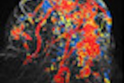
With the software, areas of enhancement that meet the minimum threshold are automatically identified as color overlays on all MRI slices. The color overlays indicate the degree of initial enhancement and allow for differentiation between persistent-, plateau-, and washout-type enhancement in the late phase after contrast injection, according to the researchers.
Six months after they initially performed manual kinetic analysis of the studies, the same two breast radiologists then read the exams with the CAD data, blinded to the initial results, treatment, and pathological outcome. In addition, two residents also interpreted the CAD datasets.
Lesions were also scored based on CAD threshold analysis alone, first using a 50% CAD threshold for initial enhancement and then a 100% threshold.
Of the 71 breast lesions included in the study, 49 were malignant, five were high-risk lesions (lobular carcinoma in situ), and 12 were benign.
Manual versus CAD techniques for breast MRI interpretation
|
The specificity gain was statistically significant (p < 0.05), although the sensitivity increase was not.
"Difference in specificity between MR interpretation ... with manual assessment of enhancement kinetics and CAD may also partly be explained by the fact that CAD provides enhancement information for all pixels in a lesion rather than for a portion of a lesion measured by using manual region-of-interest placement," the authors wrote.
Analyzing results on a per-reader basis, the first resident had 88.5% sensitivity and 93.8% specificity when using CAD, while the second resident produced 84.6% sensitivity and 81.3% specificity with the technology. The researchers did not find a statistically significant difference between either the two groups of experienced breast radiologists and residents or between any two readers separately when using CAD.
When lesions were scored based on CAD threshold enhancement alone, the software yielded sensitivity of 97.9% and specificity of 86.4% for the 50% enhancement threshold and sensitivity of 97.9% and specificity of 90% for the 100% threshold.
"With respect to interpreting the very high sensitivities reported here for CAD-based analysis based solely on the thresholding of enhancement kinetics, it should be noted that a selection bias was introduced by only including data from patients with lesions proven by core or excision biopsy," the authors noted.
By Erik L. Ridley
AuntMinnie.com staff writer
September 17, 2009
Related Reading
Fuzzy 3D ultrasound CAD sharpens breast cancer sensitivity, September 3, 2009
Breast US image analysis helps predict metastasis, July 29, 2009
Breast MRI CAD algorithm predicts cancer metastasis, February 6, 2009
Breast ultrasound CAD performance varies in ethnic populations, September 5, 2008
Breast MRI CAD doesn't improve accuracy due to poor DCIS detection, February 18, 2008
Copyright © 2009 AuntMinnie.com


