The development of biological radiation dose measurement portends a future of far greater accuracy in gauging the damage wrought by ionizing radiation in imaging exams. Two studies, focused on cardiac CT and conventional angiography exams, respectively, offer the potential of maximizing image quality while minimizing the potential radiation risk to the patient.
In back-to-back presentations at the 2009 European Congress of Radiology (ECR), investigators from the Institute of Radiology, University of Erlangen-Nürnberg, in Erlangen, Germany, and Saarland University in Saarbrücken, Germany, measured the direct in vivo effects of ionizing radiation on the double strands of blood DNA in cardiac CT and angiography exams, comparing the measurements to traditional dose-length-product and dose-area-product measurements of radiation dose.
Dr. Michael Küfner, Dr. Siegfried Schwab, and colleagues learned that sequential CT scans are less biologically damaging than spiral CT. They found that a fractionated dose delivered over time is less damaging to blood DNA than the same dose delivered all at once. And, happily, they learned that no abnormal DNA damage can be detected in the blood of interventional radiologists when measured repeatedly throughout a normal workday.
Crude dose measurements improved
Scanner readings and other dose estimates offer only the roughest indication of the actual radiation dose absorbed by the patient, Schwab explained in his ECR presentation.
"The physical dose qualities we're using day by day, and which our machines are showing us, do not adequately evaluate dose deposition in the patient -- they only say something about the dose exposition," he said. A true estimate of radiation risk requires an accurate, reproducible biological measurement of radiation-induced damage.
Recently, measuring DNA double-strand breaks (DSBs) in blood lymphocytes has emerged as a promising technique that is also easy to perform on blood withdrawn from patients before and after imaging exams.
"Previous approaches to biological dose estimation failed due to their low sensitivity," Schwab explained. "Among all the biological effects of ionizing radiation in the body, DSBs are among the most significant effects."
The exposure of mammalian cells to ionizing radiation causes DSBs within minutes. When cells are exposed, histone H2AX molecules in megabase chromatin regions adjacent to the breaks become phosphorylated. An antibody to this phosphoserine motif of human H2AX (γ-H2AX) reveals the γ-H2AX molecules appear in discrete nuclear foci that can be counted -- one per DSB.
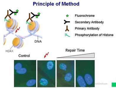 |
| Double-strand breaks (DSBs) are among the most significant DNA lesions introduced by ionizing radiation. The lesions can be visualized indirectly using foci of phosphorylated histones as a surrogate for DSBs, via γ-H2AX immunofluorescence microscopy. For example, the cell on the lower right corner of the image reveals four DSBs. All images courtesy of Dr. Siegfried Schwab. |
Thus, the researchers were able to measure phosphorylation of the histone variant that occurs after formation of DSBs in blood lymphocytes exposed to ionizing radiation. The group then used γ-H2AX immunofluorescence microscopy to count the phosphorylated histones -- again with each focus representing a single DSB within a cell.
Sequential scanning versus retrospectively gated cardiac CT
"We know that the various scan parameters have an influence on the x-ray dose in cardiac CT, but they do not integrate into the dose decision into the body of the patient," Küfner said in his presentation. On the other hand, counting DSBs provides an accurate estimate of radiation damage in vivo, he said.
"Our purpose was to determine DSB levels in patients undergoing [CT angiography (CTA)] to compare the DNA breaks produced during the spiral scan CTA and sequential CTA," he said. The study examined 32 patients, 16 on each of two scanners using various scan protocols.
Spiral images were acquired on a 64-detector-row dual-source scanner (Somatom Definition, Siemens Healthcare, Erlangen, Germany) at 100-120 kV, 330-438 mAs per rotation, and 0.2-0.39 pitch, with electrocardiogram-modulated tube current.
Sequential imaging was performed on a 128-detector-row single-source CT scanner (Somatom Definition AS, Siemens Healthcare) at 120 kV and 150-300 mA.
"We collected blood samples before and 30 minutes after the CT scan," Küfner said. The blood lymphocytes were then isolated and stained against the phosphorylated histone variant γ-H2AX, and the DSBs were visualized and counted via fluorescence microscopy.
The results showed a high correlation between dose length product (DLP) and double-strand breaks, and far greater apparent DNA damage from the spiral scans, he said.
DLP ranged from 155 to 402 mGy-cm (mean, 249) in sequential scanning and from 508 to 1,700 mGy-cm (mean, 958) in spiral scans (p = 0.00003).
The mean number of DSBs 30 minutes after coronary CT angiography ranged from 0.11 to 0.71 per cell. Total DSBs were significantly lower after sequential versus helical scans (0.14 versus 0.39 DSBs/cell, p = 0.0005). Additionally, they found that DSB levels were reduced in protocols that used 100 kV versus 120 kV; however, the difference was not statistically significant, Küfner said.
"Thirty minutes after cardiac CTA, we obtained rather high -- a mean of more than 0.39 DSBs per cell -- results in the spiral scan group," he said. "That means every second or third lymphocyte DSB is induced by the CT scan."
There was high correlation between the number of DSBs and DLP (r = 0.73), and a low (100) kV protocol led to a reduction in DSB levels, "whereas higher pitch values in patients with high heart rates is recovered by a wider pulsing window," he concluded. "Finally, a sequential scan algorithm could lead to significant reductions in DSB levels compared to patients undergoing spiral CTA."
DNA damage a function of dose, procedure time in angiography
For the second study, Schwab and colleagues (including Küfner) began with the same immunofluorescence microscopy method to detect DSBs, applying their measurements to angiography patients and their physicians, and finally validating the DSB measurements in vitro.
"The aim of the study was to detect a novel method of determining DSBs in patients undergoing angiography, and then determine the biological dose in patients and the interventional cardiologists," Schwab explained.
The radiation exposure varies widely in angiography due to changing fields-of-view during pulsed fluoroscopy/digital subtraction angiography and exposure with or without the use of contrast agents, and the process delivers fractionated irradiation over a long period of time, he said.
The researchers acquired blood samples from 37 patients undergoing angiography exams and from three interventional cardiologists before and after the examinations.
As in the previous study, the researchers visualized DSBs with immunofluorescence microscopy after staining against the phosphorylated histone variant γ-H2AX. Radiation dose to the blood was then estimated by relating in vivo numbers of DSBs to those of individual in vitro irradiated samples (50 mGy).
The results showed a dose area product (DAP) ranging from 1,337 to 12,448 µGy/m2, corresponding to fluoroscopy exam times ranging from 1.5 to 14.4 minutes.
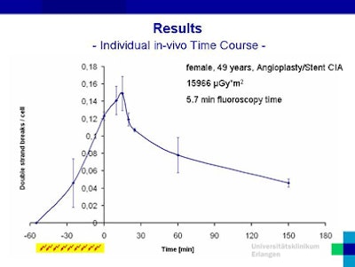
Radiation-induced increases in DSBs were found in all patients. The number of DSBs at the end of fluoroscopy ranged from 0.49 to 1.08 per cell, with the number of foci declining rapidly after the exams.
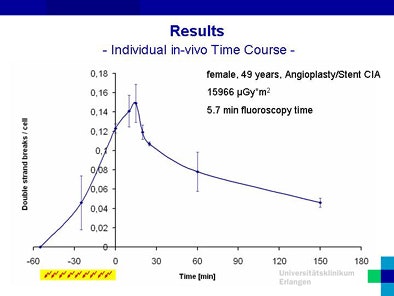 |
| DSB counts in a 49-year-old patient undergoing interventional cardiology show a spike in DNA damage following the procedure. |
"After angiography, we found high damage levels of about 0.55 [mean DSBs per cell], and at follow-up we see a reference drop in these values, which means that the DSBs are being repaired," Schwab said.
DSBs related to exam duration, anatomic region
"Dividing correlations into subgroups of different exam durations, we find moderate to very good correlations," Schwab said. "If we go one step further and normalize the DSBs per cell to the DAP, the damage levels at short examinations are much higher than after longer examinations, another effect of the repair of the DSBs."
DSB levels were also higher in abdominal exams compared to leg/pelvis exams, and they were well correlated to DAP by anatomic region (r2 = 0.497 in the abdomen).
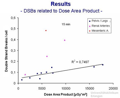 |
| DSBs versus DAP shows good correlation within anatomic regions of the exam. |
Fractionated exams less damaging than single exposure
To perform a biological dose estimation, in vitro experiments were conducted in which blood samples from healthy volunteers were irradiated at times and doses that matched those of the angiography patients, with measurement of the biological dose based on correlation of DSB levels in vivo versus DSB levels in vitro. Radiation doses to the in vitro blood samples ranged from 23.4 to 56.4 mGy.
Again, the results showed that "DSBs are repaired very quickly -- and they are repaired during angiography," he said.
Next, the blood samples were divided into two groups: one set of vials received a single dose and the other set received the same dose fractionated over 60 minutes.
"What is the effect? The maximum damage of the single-dose group was much higher than the fractioned group," Schwab said. "And the reason is that there is a mixture of repaired and newly induced DSBs.... If we had correlated the in vivo damage level to the single exposure, we would have underestimated the real blood dose" by failing to account for the repair process, he explained.
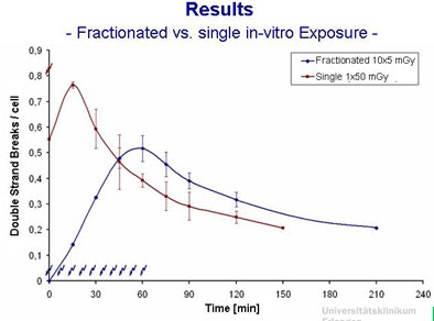 |
| In vitro results show that the same dose produces fewer DSBs when fractionated over the course of 60 minutes versus a single exposure. |
As for the multiple blood draws from three cardiologists immediately before and after performing interventional procedures, no variations were found in DSB levels -- which was certainly a relief to the interventional cardiologists, Schwab said.
"To conclude, after coronary angiography we find considerable levels of DNA damage -- up to [1.08] DSBs per cell," he said. "The number of DSBs depends on the applied dose, but it also depends on the duration and the fractionation of the exposition."
The study also demonstrated that DSB repair occurs during angiography, "so if we evaluate similar exam durations, we find the correlation of DSBs and DAP," he said. "Last but not least, during a normal working day, we could not find signs of biological effects in cardiologists."
Schwab et al's results were recently published in the German-language journal Rofo (April 2009, Vol. 181:4, pp. 374-380).
By Eric Barnes
AuntMinnie.com staff writer
May 1, 2009
Related Reading
Bisphosphonates may prevent radiation-induced leukemia, April 21, 2009
Researchers find genes that affect radiation damage, April 9, 2009
Higher cancer risk seen in frequently scanned patients, March 31, 2009
Morning radiation therapy minimizes side effects for some patients, February 26, 2009
Prenatal radiography does not increase risk of childhood brain tumors, December 31, 2007
Copyright © 2009 AuntMinnie.com


