Radiology departments have been in the forefront of implementation of technology within healthcare, and are poised to be so yet again with artificial intelligence (AI). In the coming years, PACS with AI will replace PACS without AI.
Thanks to the proliferation of speech recognition technology, most radiology departments already have extensive practical experience with AI. Speech recognition software is based on a form of AI called computer audition, which mimics the function of the human hearing system. An audio clip is passed through an artificial neural network and text is produced.
Speech recognition uses machine learning and deep learning with multiple neural networks. The widespread adoption of this form of AI is due to integration of speech recognition into radiology reporting workflows. RIS and teleradiology platforms are commonly deployed in the U.K.'s National Health Service (NHS) for reporting workflow, and speech recognition has been adopted across Europe, including all NHS trusts (hospital groups).
Computer vision
In contrast to computer-audition AI, computer-vision AI deals with digital images. It automates the tasks of the human visual system to process, analyze, and understand digital images to produce symbolic and numerical data.
 Dr. Neelam Dugar is a consultant radiologist at the Doncaster & Bassetlaw Hospitals NHS Trust, U.K., and informatics adviser to the Royal College of Radiologists.
Computer-aided detection (CAD) software has been around for many years. However, with the advent of machine learning and deep learning, there is huge potential for improved detection accuracy as well as better specificity.
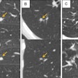
CAD software can support radiologists in detecting subtle lesions early. As part of the radiology reporting workflow, radiologists expect that CAD markers will be superimposed on images and can be toggled on and off. Some radiologists may like the CAD markers to be displayed during primary review of images, while others may like it displayed on-click. This allows radiologists to use computer-vision AI as the second reader for detection.
A large amount of research continues to show that computer vision is more sensitive than human vision. However, the interpretational skills of radiologists are still required to determine, for example, if a computer-detected anomaly is "artifactual" or real, or if the anomaly is related to infection or tumor.
Interpretation of images to provide a tentative and differential diagnosis relies upon the radiologist's experience, analysis of the clinical history, blood results, histopathology reports, and other clinical information available in the electronic patient record.
Anomaly, change detection
Computer-vision AI comes in two forms: anomaly detection and change detection.
Anomaly detection AI provides markups to assist reporting or raising the priority of reporting. Mammography shadow CAD, CT lung nodule CAD, CT brain anomaly CAD, chest x-ray shadow CAD, and plain x-ray limb fracture CAD are examples of anomaly detection AI that are of interest to most radiologists.
The other form of computer-vision AI -- change detection -- allows automatic registration of the baseline and follow-up images with linked cursors. It allows radiologists to toggle between baseline and follow-up images with color-coded change maps. This can be used for change detection in follow-up of tumors, multiple sclerosis, and brain infarcts.
Levels of computer vision
The three levels of computer-vision AI include the following:
- Simple computer detection followed by interpretation by radiologists
- Computer detection with decision support (suggestion of possible diagnosis due to analysis of image features) followed by human interpretation
- Independent computer diagnosis without human interpretation
Due to the complexity of medicine and medical sciences, the third level of AI seems rather distant at the moment. There are various ethical hurdles to be crossed to reach the third level of AI, which is devoid of human judgement. Huge numbers of validated datasets of various disease processes will be needed to pass any ethical approval.
The first and second levels of computer-vision AI will certainly enhance and augment human interpretation by radiologists. In addition, the barriers for entry to these first two levels are much lower than the third, but if this technology is to be useful, it needs to be integrated into our PACS reading workflows.
It is well known there are huge volumes of reporting backlogs within NHS radiology departments. There is always the risk there will be a few patients who have lung cancer within the chest x-ray reporting backlog. A delay of up to one month is possible before a radiologist can report the chest x-ray.
The use of computer-vision AI could transform prioritization within the backlog reporting by facilitating computer-aided simple triage. When an anomaly is detected by the computer-vision AI software within the reporting backlogs, this exam could be prioritized within the radiologist reporting worklists. This would reduce the risk of delayed diagnosis.
However, this requires vendor cooperation around HL7 standards. The system providing computer-vision AI should be able to update the HL7 order-entry message to raise the priority of reporting the abnormal study within reporting worklists.
There are a large number of companies already providing computer-vision AI software for anomaly detection and change detection. All these forms of computer-vision AI are appearing in various articles, and are being developed by both small and large vendors.
Limited real-world availability
Whilst computer-vision AI is developing at a very rapid pace in the commercial world, the availability in the real world for radiologists is very limited. PACS companies have failed to integrate computer-vision AI into their systems. I do not know of any PACS company that includes CAD software within their PACS offerings for mammography CAD, CT lung nodule CAD, brain CT anomaly CAD, chest x-ray shadow detection CAD, and fracture detection CAD.
Similarly, no PACS company is integrating computer-generated change detection for tumor follow-up or multiple sclerosis follow-up in their offerings. Radiology departments and radiologists around the world desperately need CAD AI to supplement and augment their reporting in the above areas as a minimum, but this needs to be integrated into existing PACS software using DICOM CAD standards.
If integrated within the radiologist reporting workflow, computer-vision AI has the potential to transform the efficiency and accuracy of radiology reports. As PACS customers become more discerning, it is highly likely that artificially intelligent PACS will replace PACS that are not artificially intelligent.
Content aggregation
Also worth a mention is another form of AI called content-aggregation AI, which could be programmed to extract information from diverse healthcare systems and then display this information for the radiologist. To make accurate clinical diagnoses, radiologists need more clinical information, including blood test results such as liver function tests, tumor markers, renal function tests, blood amylase levels, and inflammatory markers.
Radiologists also need access to histopathology reports, endoscopy reports, and other clinical documents and images. Context-aggregation AI adoption would enable content aggregation from healthcare data silos. PACS companies using AI need to be at the forefront of enabling content aggregation to help radiologists make better and more accurate diagnoses.
Content aggregation and computer vision are the forms of AI that are most needed by radiologists to make them more efficient and smarter. PACS vendors must study the market closely, talk to visionary radiologists, and then integrate computer vision and content aggregation AI into PACS software.
Don't get left behind. PACS with AI will certainly replace PACS without AI in the next five to 10 years.
Dr. Neelam Dugar is a consultant radiologist at the Doncaster & Bassetlaw Hospitals NHS Trust, U.K., and informatics adviser to the Royal College of Radiologists (RCR). This article was written in her personal capacity, and her views and ideas are not necessarily shared by the RCR.
The comments and observations expressed herein do not necessarily reflect the opinions of AuntMinnieEurope.com, nor should they be construed as an endorsement or admonishment of any particular vendor, analyst, industry consultant, or consulting group.


