In 2016, Geoff Hinton, father of artificial intelligence (AI) and machine learning (ML), caught the attention of politicians by declaring that AI and ML would replace radiologists. He couldn't have been further from the truth. It is clear from all of the recent presentations at ECR and RSNA that radiologists will become smarter and more efficient with the help of AI, but they will not be replaced.
There are many types of computer vision algorithms being assessed for use in radiology. The following are some examples:
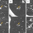
- Computer-aided anomaly detection: Algorithms would be used for detection of abnormalities such as breast lesions, lung nodules on CT, lung shadows on chest x-ray, stroke on CT, hemorrhage on CT, fracture on plain x-ray, colonic polyp, and pulmonary embolism detection on CT.
- Computer-aided simple triage: When an anomaly is detected, algorithms could be used to raise the priority of reporting in the worklist. This is being used for detection of hemorrhage in CT head scans and prioritizes reporting.
- Computer-aided change detection: Computer algorithms would detect change on serial studies -- for example, multiple sclerosis, tumor progression, etc.
- Image fusion or coregistration: These algorithms allow fusion of images between different modalities, including CT and MRI.
- Computer-aided classification: This is often called computer-aided diagnosis. It provides a possible diagnosis/nature of the lesion or malignancy risk. Algorithms take into account image features, location of lesion, and radiomics (textural analysis by computer that is not detected by the human eye) to assess the risk of malignancy or the likely diagnosis. It is being used for assessing risk of malignancy in breast and lung lesions.
- Computer-aided segmentation: Algorithms are used for drawing around structures on the images. It can be used for drawing around tumors or even around normal structures for radiotherapy planning. It can also be used for display of an angiogram from a CT or MRI exam.
- Computer-aided quantification: Algorithms will count the number, size, and volume of abnormalities such as lung nodules on CT. It can be used for assessing percentage involvement of lungs in interstitial lung diseases, quantification of bone age, and degree of vessel stenosis. It can provide an emphysema index, osteoporosis score, and calcium score.
- Computer-aided prediction: Algorithms will be used for predicting the onset of stroke, prognosis of disease, survival, and response to therapy. For instance, by quantifying the shape, volume, and texture of the hippocampus, algorithms may be used for predict the risk of dementia.
Diagnostic accuracy of algorithms
 Dr. Neelam Dugar is a consultant radiologist at the Doncaster & Bassetlaw Hospitals NHS Trust, U.K., and informatics adviser to the Royal College of Radiologists.
It is really important for us to know how much we really can rely on AI. AI is a diagnostic tool, and like any diagnostic test, AI algorithms have both a sensitivity and specificity. No algorithm can claim to be 100% accurate. Hence, it is really important that when doctors take decisions on management for the patient, or radiologists issue a narrative actionable report, they are aware of the limitations. For example, the sensitivity and specificity of the algorithm used for defining risk of breast malignancy could be 95% sensitive and 80% specific. This encourages doctors to review the images themselves when offering advice or issuing a narrative actionable report.
Display of sensitivity and specificity of AI algorithms for the frontline doctors and reporting radiologist is essential for patient safety. When the output of computer vision AI is sent to a PACS or enterprise viewer, it should be mandatory that the sensitivity and specificity of the algorithm used are displayed along with the images and CAD/AI markers.
Factors on which accuracy depends
The accuracy of these algorithms is dependent on two important factors: the type of algorithms used and also the acquisition parameters applied by the modality. If the algorithm is to be accurate, it is really important the acquisition parameters are standardized prior to application of the algorithm. Hence, many of the computer vision AI algorithms need to be integrated with the modality. For example, if there is a requirement for lung nodule detection on CT chest protocol, the scanner should automatically reset the acquisition parameters to optimize lung nodule detection.
Artificially intelligent machines and scanners
Current x-ray machines, CT scanners, and MRI scanners produce images only. In the future, these scanners will be replaced by intelligent machines. All digital radiography units will apply a chest shadow algorithm and produce images along with markers for shadows. The sensitivity and specificity of the detection marker will be displayed along with the images. Similarly, digital radiography machines will have fracture analysis algorithms. The sensitivity and specificity of the output will be sent to PACS along with fracture markers. Similarly, CT scanners will have a lung nodule detection algorithm applied. CT scanners will also output the emphysema index, osteoporosis index, and calcium score along with their sensitivity and specificity for calculation for each of these algorithms.
Artificially intelligent PACS
Currently, PACS is only capable of displaying images as sent by modalities. However, in the future, they will be expected to display CAD markers as well. PACS vendors will also apply some computer vision algorithms. These include image fusion or coregistration. This will enable radiologists to fuse images from different modalities while automatically modifying the zoom, slice thickness, and the acquisition parameters. PACS vendors may apply computer-aided segmentation algorithms for anomalies such as liver metastasis (for comparison and follow-up).
PACS reading workflow
At present, radiologists are not supported by computer vision AI. However, the rapid pace of development is afoot, and the radiology reporting workflow is going to change.
Currently, radiologists read images without any CAD markers. In the future, many forms of detection markers along with quantification and classification of anomalies will support radiologists' reading workflow. We will need to learn how to issue narrative actionable reports in the context of AI while understanding the sensitivity and specificity of the algorithms applied. We will need to be able to discount anomalies when the computer gets it wrong. We will need to provide more human understandable narrative reports from the scientific jargon provided by computer algorithms.
Once again, radiologists' working patterns are about to change enormously. Computer vision will help radiologists detect subtle masses on mammograms, detect lung nodules on CT, fractures on plain x-ray, shadows on chest x-ray, etc. Computer vision will also provide a malignancy index for breast lesions and lung nodules. Outputs such as emphysema index, osteoporosis score, and calcium score will be generated by CT scanners.
Computer vision will aid radiologists in producing narrative and actionable reports. These computer vision algorithms will provide a huge amount of decision support for radiologist reporting. It is envisaged that the modalities such as digital radiography units, CT systems, and MRI scanners will output intelligent information along with images. Intelligent machines will replace the current machines. It is also envisaged that current PACS will be replaced by intelligent PACS. Radiologists will learn to understand these computer-generated scientific outputs to produce human-readable actionable reports to support patient management decisions. Find out more in the U.K. Royal College of Radiologists' guidelines here.
Research and reality
It is clear that computer scientists are busy at work creating a myriad of algorithms. Given the success of facial recognition technology in passport scanning systems, Facebook, etc., it is not surprising that computer vision is being applied successfully to radiology to detect lung shadows, nodules, fractures, hemorrhages, etc. However, very few algorithms have appeared yet in the real world to support clinical working due to stringent regulations.
Recently, there has been a reclassification of CAD software by the U.S. Food and Drug Administration (FDA) from class III to class II. This may encourage more algorithms to appear in clinical practice, and that will be a very welcome change for patients and doctors.
Dr. Neelam Dugar is a consultant radiologist at the Doncaster & Bassetlaw Hospitals NHS Trust, U.K., and informatics adviser to the Royal College of Radiologists (RCR). This article was written in her personal capacity, and her views and ideas are not necessarily shared by the RCR.
The comments and observations expressed herein do not necessarily reflect the opinions of AuntMinnieEurope.com, nor should they be construed as an endorsement or admonishment of any particular vendor, analyst, industry consultant, or consulting group.


