PET imaging biomarkers have become central to understanding and diagnosing cases of dementia and Parkinson's disease, but it's vital to keep in mind the main pitfalls and artifacts, according to Spanish researchers.
"PET imaging allows for the in vivo quantification of molecular targets with high sensitivity, aiding in the study of disease pathophysiology and progression from preclinical stages," Angela Bronte, a doctoral candidate at Clínica Universidad de Navarra, Madrid, and colleagues wrote in an article posted on 6 February by European Radiology. "By visualizing specific molecular pathologies, PET biomarkers enable a shift from symptom-based to biology-based definitions of neurodegenerative diseases, allowing for earlier and more accurate detection and diagnosis. This has significant implications for developing and testing new therapies aimed at modifying disease course."
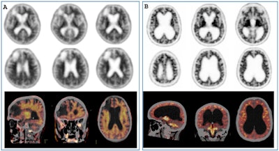
 Pitfalls and artifacts in amyloid PET imaging. (A) F-18 florbetapir PET/CT scan of a patient with right frontal stroke and brain atrophy. The PET/CT fusion images clearly show no difference between white matter and grey matter (positive amyloid PET scan). (B) F-18 florbetapir PET/CT scan of a patient with normal pressure hydrocephalus. In this case, the fusion images showed much higher activity in the white matter than in the grey matter (negative amyloid PET scan). All figures courtesy of Angela Bronte, Prof. Javier Arbizu, PhD, et al and European Radiology.
PET molecular imaging tools for the clinical diagnosis of patients presenting with cognitive impairment or parkinsonism and suspected neurodegenerative disease vary widely in their image acquisition protocols and image interpretation, but standardized visual reading methods and specific semiquantitative parameters help greatly, they explained.
The current trend in defining clinical diagnostic criteria for neurodegenerative diseases is toward a more biological approach, focusing on molecular and cellular mechanisms rather than clinical symptoms alone. Diseases are increasingly being characterized as so-called proteinopathies, with specific protein aggregates serving as hallmarks for different conditions. As a result, there is an increasing focus on identifying biomarkers for early diagnosis and monitoring disease progression, particularly as new disease-modifying treatments become available, the authors pointed out.
![(A) [F-18] FDOPA PET image of a healthy patient (A.1) and a patient with Parkinson's disease (A.2). B: Pitfalls and artifacts in presynaptic dopaminergic imaging. [F-18F] FDOPA PET scan (B.1) of a patient with suspected Parkinson's disease showing a decrease in the right putamen. However, the PET/MRI image (B.2) shows that the decreases are due to the artifact of spaces surrounding the walls of vessels within the brain parenchyma observed on MRI.](https://img.auntminnieeurope.com/files/base/smg/all/image/2025/02/2025_02_13_mol_insider_Figure_6.67ad30a73f2ff.png?auto=format%2Ccompress&fit=max&q=70&w=400) (A) [F-18] FDOPA PET image of a healthy patient (A.1) and a patient with Parkinson's disease (A.2). B: Pitfalls and artifacts in presynaptic dopaminergic imaging. [F-18F] FDOPA PET scan (B.1) of a patient with suspected Parkinson's disease showing a decrease in the right putamen. However, the PET/MRI image (B.2) shows that the decreases are due to the artifact of spaces surrounding the walls of vessels within the brain parenchyma observed on MRI.
(A) [F-18] FDOPA PET image of a healthy patient (A.1) and a patient with Parkinson's disease (A.2). B: Pitfalls and artifacts in presynaptic dopaminergic imaging. [F-18F] FDOPA PET scan (B.1) of a patient with suspected Parkinson's disease showing a decrease in the right putamen. However, the PET/MRI image (B.2) shows that the decreases are due to the artifact of spaces surrounding the walls of vessels within the brain parenchyma observed on MRI.
"PET tracers that bind to beta-amyloid plaques, particularly currently available fluorinated tracers, enable the detection of amyloid pathology years before clinical symptoms of Alzheimer's disease appear as it is one of the main neuropathological hallmarks, although it is not specific," they continued.
Amyloid PET can identify individuals in the preclinical stages of Alzheimer's disease, allowing for earlier intervention and recruitment into clinical trials. Also, newer tau-PET tracers can visualize neurofibrillary tangles -- another hallmark of Alzheimer's pathology. Tau PET can track disease progression and neurodegeneration more closely than amyloid imaging, but the most available tracer worldwide is FDG-PET, which measures glucose metabolism and detects subtle changes in brain function before structural changes are apparent on MRI, the researchers added. Characteristic patterns of hypometabolism can be seen in the early stages of various neurodegenerative diseases.
![Pitfalls and artifacts in [F-18] FDG PET imaging. (A) Scan of a patient with central nervous system drug interference shows cortical hypometabolism that could mimic an AD pattern, but with thalamic hypometabolism characteristic of this interference. (B) Scan of a patient with a right frontoparietal ischemic antecedent (arrow), showing a diaschisis of the contralateral cerebellum (arrow) due to disruption of the corticospinal tract. (C) An example of a PET scan with motion artifacts.](https://img.auntminnieeurope.com/files/base/smg/all/image/2025/02/2025_02_13_mol_insider_Figure_5.67ad30bfe2686.png?auto=format%2Ccompress&fit=max&q=70&w=400) Pitfalls and artifacts in [F-18] FDG PET imaging. (A) Scan of a patient with central nervous system drug interference shows cortical hypometabolism that could mimic an AD pattern, but with thalamic hypometabolism characteristic of this interference. (B) Scan of a patient with a right frontoparietal ischemic antecedent (arrow), showing a diaschisis of the contralateral cerebellum (arrow) due to disruption of the corticospinal tract. (C) An example of a PET scan with motion artifacts.
Pitfalls and artifacts in [F-18] FDG PET imaging. (A) Scan of a patient with central nervous system drug interference shows cortical hypometabolism that could mimic an AD pattern, but with thalamic hypometabolism characteristic of this interference. (B) Scan of a patient with a right frontoparietal ischemic antecedent (arrow), showing a diaschisis of the contralateral cerebellum (arrow) due to disruption of the corticospinal tract. (C) An example of a PET scan with motion artifacts.
"Evaluation of neuronal activity and neurodegeneration by means of [F-18] FDG PET, a worldwide highly available technique included in the majority of clinical diagnostic criteria of neurodegenerative diseases, requires proper patient preparation, as well as PET facility and personnel training," stated Bronte, who now works in the nuclear medicine department at the Hospital Universitario de Son Espases, Palma de Mallorca.
Future research
The Madrid team is now working on some additional projects related to neuroimaging, including the following:
- Assessment of four-repeat (4R) tau deposition in the brain with F-18 PI2620 in progressive supranuclear palsy (PSP) compared with controls without 4R neurodegenerative tauopathy: a clinical trial. "In this study, we aim to define in vivo the correlations between tau deposits and the pattern of neurodegeneration detected by PET in patients with different clinical PSP subtypes," Prof. Javier Arbizu, PhD, chair of nuclear medicine at the University of Navarra told AuntMinnieEurope.com on 13 February. "We are performing an in vivo cross-sectional study of patients with clinical diagnosis of PSP and controls who will undergo a tau-PET scan using F-18 PI2620 and glucose metabolism PET using F-18 FDG, as well as brain MRI and clinical and neuropsychological evaluations.
- A neuropathological study on postmortem tissue from neuropathologically confirmed PSP and Parkinson's disease cases available in the Brain Bank of Navarrabiomed is being conducted in order to analyze tau load and distribution by immunohistochemistry techniques. The data will be correlated with the expression of F-18 PI2620 in the same samples by means of autoradiography.
- Analysis of Amyloid PET images with Centiloid scale: investigation of the robustness of the technique and impact in clinical diagnosis in equivocal cases.
- Retrospective analysis of the impact of FDG-PET imaging in the diagnostic evaluation of patients with autoimmune encephalitis: a multicenter study.
- Limbic-predominant age-related TDP-43 encephalopathy (LATE)-like pattern in FDG-PET of mild cognitive impairment (MCI) subjects: clinical characteristics, amyloid burden, and conversion to Alzheimer's disease.
You can read the full European Radiology paper here. The co-authors were Elena Prieto, Gemma Quincoces, Elena Erro, and Javier Arbizu.


