Using photon-counting CT (PCCT) for abdominal imaging to diagnose kidney stones reduces patients' radiation exposure by 44% compared with conventional CT, researchers have found.
PCCT also offers a higher signal-to-noise ratio than conventional CT with no evidence of a difference in reader confidence, reported a team led by Dr. Henner Huflage of University Hospital Würzburg in Germany. The findings were published on 30 July in Radiology.
Urolithiasis is a common disease with increasing prevalence among adults and even children in recent years, and since it can recur, there's often frequent need for diagnostic imaging, the group explained. Noncontrast, low-dose CT of the abdomen is the go-to exam for the diagnosis of kidney stones, with a sensitivity rate of 95% and a specificity rate of 98%. But complying with the American College of Radiology (ACR) guideline of radiation exposure rates of no more than 3 mSv for low-dose CT can be tricky.
That's where PCCT comes in. Previous studies have shown it can produce images without electronic noise, improved tissue characterization, and optimized spectral imaging while also using less iodine and radiation. But how does it compare with traditional CT for kidney stone diagnosis?
To address the question, Huflage and colleagues compared radiation dose and image quality of energy-integrating detector (EID) CT with those of an experimental PCCT scan protocol in patients suspected of having kidney stones. The study included 507 patients who underwent unenhanced abdominal PCCT (229 patients) or EID CT (278 patients) for the indication between February 2022 and March 2023. The researchers evaluated signal-to-noise results at the kidney, psoas, and obturator muscle; and three radiologists assessed the presence of kidney stones and registered their diagnostic confidence using a five-point scale (1, little-to-no confidence; 5, complete confidence).
The group reported an effective radiation dose for EID CT of 1.39 mSv and for PCCT of 0.79 mSv, a reduction of 44%. Volume CT dose index was 1.99 mGy for EID CT and 1.17 mGy for PCCT, a reduction of 41%. The researchers also found the following:
| Performance comparison of EID CT and PCCT for detecting kidney stones on abdominal imaging | ||||
|---|---|---|---|---|
| Measure | EID CT | PCCT | Percent difference | p-value |
| Image noise (in Hounsfield units) | ||||
| Kidney | 21.24 | 15.88 | 25% | < 0.001 |
| Psoas muscle | 21.71 | 16.34 | 25% | < 0.001 |
| Internal obturator muscle | 19.61 | 15.61 | 20% | < 0.001 |
| Signal-to-noise ratio | ||||
| Kidney | 1.66 | 2.15 | 30% | < 0.001 |
| Psoas muscle | 153 | 114 | 23% | < 0.001 |
| Internal obturator muscle | 2.79 | 3.25 | 17% | < 0.001 |
The authors noted that agreement among the three readers was "almost perfect" (Krippendorff's alpha coefficient, ≥ 0.99) and diagnostic confidence ratings had "good reliability" (with a correlation coefficient of 0.78 [1 as reference]).
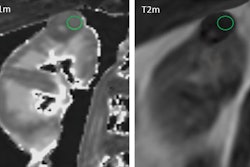
 Axial contrast-unenhanced abdominal CT examinations in a 38-year-old female patient with a body mass index (calculated as weight in kilograms divided by height in meters squared) of 31.23 with known renal calculi for recurring events of flank pain. The patient underwent (A) dose-optimized energy-integrating detector CT (1.49 mSv) and (B) submillisievert photon-counting detector CT (0.94 mSv). Images from both examinations showed a 3-mm calculus in the left kidney (arrows). Images and caption courtesy of the RSNA.
The study findings are promising, according to the authors.
"[Our study showed that] the radiation burden could be reduced by an additional 44% compared with that of fully optimized EID CT with an established low-dose protocol while maintaining reader confidence and even increasing image quality," they wrote.
Dr. Nariman Nezami of Georgetown University Medical Center in Washington, DC, and Dr. Ashkan A. Malayeri of the National Institutes of Health in Bethesda, MD, agreed in an accompanying editorial.
"As radiologists, we strive to enhance the quality of patient care and minimize risk," they wrote. "[This study] shows the potential of [PCCT] in increasing diagnostic confidence in renal stone depiction, improving image quality, and decreasing radiation dose, marking an important advancement toward this goal."
The complete study can be found here.