Radiologists in Turkey have described events that occurred in the first 24 hours in a radiology unit after two major earthquakes devastated the country on 6 February 2023 and provided details on the imaging exams performed.
The first earthquake occurred at 4:17 a.m., noted Dr. Mehtap Ilgar, of Ankara Etlik City Hospital in Ankara, and Dr. Nurullah Dağ of Malatya Training and Research Hospital, a large trauma center 50 miles from the epicenter. Only one radiologist and seven radiology technicians were on duty at the time.
“Within half an hour, the patient load increased, and all available radiology personnel were called to emergency duty,” the authors wrote, in an article published on 22 August in Tomography.
The epicenter of the second earthquake was about 80 miles away and occurred at 1:24 p.m. local time. Radiology staff who were at their homes during the first earthquake worried about their families, the authors wrote. Meanwhile, aftershocks continued, and the anxiety and fear of the building collapsing increased, they wrote.
“The second earthquake shows that worst-case scenarios should always be considered when planning for disasters, the authors wrote.
More than 50,000 people died, and more than 100,000 people were injured in the earthquakes. In all, over a 24-hour period, the unit in Malatya performed imaging for 596 patients. Five x-ray units, two 128-detector CT units, one 16-detector CT unit, and portable ultrasound equipment were available.
Despite a power outage, the generators were on and the imaging equipment was working, the authors wrote. They said they had an advantage in that their x-ray and CT machines were located on the ground floor, as some of the elevators were damaged. In addition, the large aftershocks made use of the elevators risky, they wrote.
 The number of patients affected by region. Images available for republishing under Creative Commons license (CC BY 4.0 DEED, Attribution 4.0 International) and courtesy of Tomography.
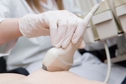
Ultrasound and CT reports were handwritten on paper or verbally communicated by radiologists to the physicians face-to-face in the emergency department. Within the first six hours, ultrasound was the most useful and accessible modality, yet there were no suitable computers nearby where radiologists could enter these reports, they noted.
In total, 437 (73.3%) out of 596 patients underwent CT scans, a majority being whole-body CT without contrast unless clinically indicated. The most commonly affected regions were the thorax, vertebrae, and extremities.
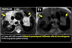
Contusions were most common in patients with thoracic findings. Contusions were seen in 35 (67.3%) patients, rib fractures in 21 (40.4%), pneumothorax in 20 (38.5%), hemothorax in 18 (34.6%), and laceration in three patients (5.8%). In 26 (50%) patients, multiple thoracic findings were seen together.
The most common finding was fractures. Out of 160 patients with pathological findings, 139 (86.9%) had evidence of fractures. Fractures were observed in a single bone in 84 (60.4%) patients and in more than one bone in 55 (39.6%) patients. The fibula, femur, and tibia were the bones with the most frequent fractures.
“When developing disaster preparedness plans, radiology departments should be actively involved to ensure their prompt and effective operation,” the authors concluded.