Important hurdles such as reproducibility and lack of automation must be overcome before radiomics is integrated as a clinical adjunct in ovarian cancer detection, but artificial intelligence shows considerable promise in this area, according to a leading European specialist in women's imaging.
"Integration of artificial intelligence techniques will not only assist in solving these [hurdles] but likely provide new prognostic algorithms for patient-tailored therapies," noted Dr. Rosemarie Forstner, associate professor of radiology at Salzburg University Hospital in Austria. "Radiomics and radiogenomics potentially offer a broad spectrum of complementary information in ovarian cancer diagnosis and treatment."
The combination of radiomic features and clinical data may allow for the creation of predictive models of resectability or of tumor progression, she explained in an editorial posted on 28 May by European Radiology.
Despite advances in therapy, ovarian cancer remains a major challenge. Only a marginal improvement in overall survival has occurred, largely because ovarian cancer is mostly diagnosed late and subsequently will relapse, she pointed out. In contrast, borderline tumors and stage I invasive ovarian cancer have excellent prognoses.
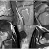
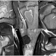
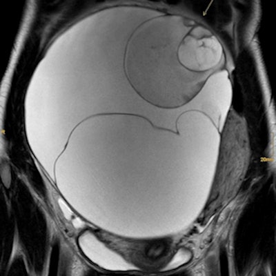
 Mucinous borderline tumor and stage IA invasive ovarian cancer in a 28-year-old woman. Coronal T2 (a) demonstrates a large multilocular cystic mass of the right ovary typical of a mucinous tumor. At its superior aspect areas with irregular septations, contrast enhancement (b), and restricted diffusion (c) are demonstrated (arrow).
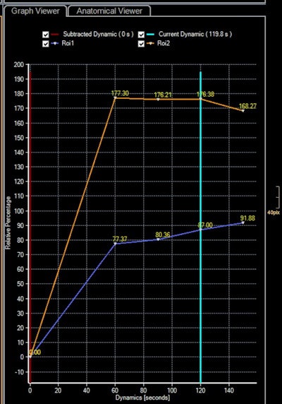 Time-intensity curves of the uterus (orange) and solid tissue of the mass (blue) demonstrate type 2 curve with typical initial rise followed by a plateau. At histopathology, in this area, foci of invasive cancer were seen. Images courtesy of European Radiology.
Early detection remains one of the unmet needs in the management of this disease. Transvaginal sonography and MRI are effective tools to characterize ovarian masses, and greater standardization of imaging technique and implementation of predictive models of risk of malignancy can contribute to better detection, Forstner added.
"The ability to detect ovarian cancer before it metastasizes is crucial, as borderline tumors and most stage I invasive cancers have a 5-year survival rate of more than 90%," she wrote. "Furthermore, accurate preoperative assessment may allow a tailored patient-centered approach for adnexal masses when these are assessed by ultrasound or in indeterminate cases complemented by MRI."
Preoperatively identifying lesions as benign can prevent unnecessary surgery or allow minimally invasive approaches and reduce patient anxiety, whereas in suspected malignancy or indeterminate findings, referral to specialist cancer facilities will guarantee adequate treatment.
Development of biomarkers
No epigenetic biomarkers are available for the early detection of ovarian cancer from tissues or fluids, but gene expression and methylation-based arrays and other emerging techniques such as liquid biopsies or autoantibody serum biomarkers are under development, Forstner continued.
"Radiomics and radiogenomics from CT or MRI data have opened new insights in ovarian cancer tumor biology," she stated. "New, rapidly evolving applications include identification of radiomic features and their correlation with phenotype, genetic features, and prediction of cancer progression or prognosis."
The key advantage of radiomics in highly heterogeneous tumors such as high-grade serous ovarian cancer is the evaluation of the whole tumor/tumor burden that is impossible at a biopsy level. In this setting, a radiomic approach may allow the characterization and quantification of inter- and intratumoral heterogeneity linked with prognosis and drug resistance, she concluded.


