Australians excel at numerous sports, but what's their approach to sports imaging? What injuries do radiologists see and how do they diagnose and treat them? Dr. David Connell, a musculoskeletal (MSK) radiologist and the clinical director at the Imaging Olympic Park in Melbourne, speaks about his philosophy and daily work.
Connell is an associate professor at the Monash University Faculty of Medicine and also at the faculty of sports medicine and research at La Trobe University in Melbourne. He is a past president of the Australasian MSK Imaging Group and was a radiologist at the Sydney and London Olympics.
European Society of Radiology: Sports imaging is the main theme of the International Day of Radiology (IDoR) on 8 November. In Australia, is there a special focus on sports imaging within radiology training or special courses for radiologists?
 Dr. David Connell.
Connell: Radiology training takes place in public hospitals, which, other than trauma, do not usually see injuries in the sporting population. Athletes are most commonly treated at private MSK centers, and it is in this setting that interested radiologists may gain exposure at advanced training/fellowship positions.
Please describe your regular working environment and schedule.
I work in a dedicated musculoskeletal practice located in the bowels of a rugby and soccer stadium. Sports-related imaging and intervention make up 100% of our work. Our facility is the premier sports imaging center in Australia. We take on two fellows per year, and the knowledge they acquire can be disseminated throughout the country. One exercise allocated to the fellow is a "case of the week," which involves describing an interesting case, explaining the anatomy, pathophysiology, and literature review. This case is emailed to more than 2,000 doctors per week. We also hold regular clinical meetings and review sessions.
Which sports produce the most injuries that require medical imaging? Have you seen any changes here?
AFL (Australian Football League) football, rugby, and netball account for most injuries -- much more than, say, soccer, basketball, and cricket. As the physical demands of these sports have changed, we see many more muscle injuries (particularly hamstring and calf) than before. In the last five years, we have seen more women transition over to football and rugby. As a consequence, we are seeing many more injuries in the female population than before; these include muscle tears, shoulder dislocations, and anterior cruciate ligament (ACL) rupture. The incidence of ACL rupture in AFL football for women is nine times that of the male population, which is quite worrisome.
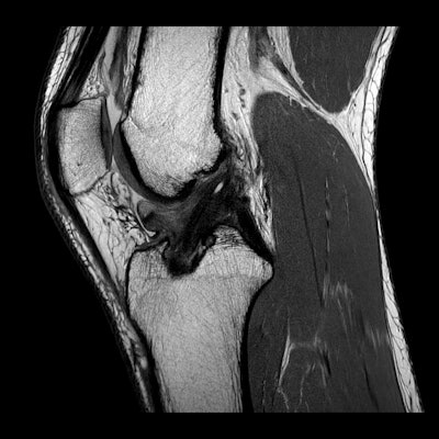 A 28-year-old female professional football player with a past history of ACL rupture of the right knee presents with anterior knee pain and locking. Top: Sagittal MR image shows wad of arthrofibrosis in the intercondylar notch. Bottom: Axial MR image shows ovoid focus of scar tissue in anterior aspect of notch. All images courtesy of Dr. David Connell.
Please give an overview of the sports injuries with which you are most familiar and their respective modalities.
As a diagnostic tool, MRI has superseded both ultrasound and radiographs. For example, we typically perform 80 musculoskeletal MRI scans per day but perhaps fewer than 10 radiographs. There is a broad mix of all the joints. We are now quite confident in assessing muscle injuries and providing accurate information about when an athlete might return to play and the risk of a recurring injury. For intervention, fluoroscopy has become redundant, and we would typically perform 50 to 60 interventions per day under ultrasound or CT guidance.
Our athletes are very rarely subject to radiation exposure, simply because almost all diagnostic imaging is performed with MRI or ultrasound. Likewise, a lot of interventions are performed with ultrasound. Even when we perform CT-guided injections for backs, our scanners have a very low radiation dosage.
What's the role of the radiologist within the clinical team?
Radiologists are able to provide an objective measure of a sporting injury. This has to be fed into the team and assessed in the context of clinical parameters. I often hear sports doctors saying that imaging is unnecessary or doesn't change management -- but I have yet to come across one brave enough to treat an elite athlete without an imaging assessment. Having said that, radiologists are a small cog in the machine, and it is important to remember to always treat the patient, not the scan.
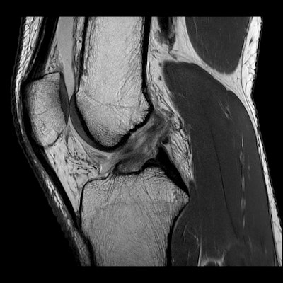 Same 28-year-old female football player with acute pivot shift injury. Top: Sagittal MR image shows failure of the native ACL injury. Bottom: Corresponding fat-suppressed sagittal image shows contusion beneath the posterior lip of the lateral tibial plateau.
Imaging can detect almost all significant injuries in sport. When imaging is negative, this is most reassuring to the athlete and club that nothing terrible has occurred. This is particularly the case with head injury/concussion and muscle injuries.
How much involvement do you have in treatment and follow-up?
Our center is a diagnostic and treatment facility for sporting injuries. We undertake diagnostic imaging, and then liaise with the referring sports clinician, surgeon, and player. In many cases, we proceed to an imaging-guided intervention. We often have the player returning for follow-up imaging to assess for healing of an injury and provide valuable information about return to play.
How can imaging be used to prevent sports-related injuries and diseases? Can the information provided by imaging be used to enhance the performance of athletes?
We are often asked to screen patients prior to the commencement of a season. For example, we have undertaken preseason ultrasound assessments of the patellar and Achilles tendons of entire football teams. The outcome is fascinating, as often asymptomatic players can be found with abnormal imaging findings. These players have a higher risk of developing symptoms in the course of the season; their training and load can be modulated accordingly to prevent injury.
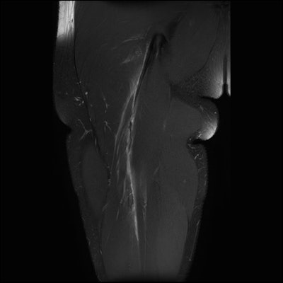 A 25-year-old professional football player presents with an acute injury to the right hamstring. Top: Coronal fat-suppressed image shows rupture of the intramuscular tendon rachis of the biceps femoris muscle in the proximal third of the thigh. Note detensioning of the intramuscular tendon, resulting in a wavy appearance. Bottom: Corresponding axial scan shows absence of the intramuscular tendon as consequence of tendon failure and retraction.
Has the demand for imaging increased in sports medicine? What can be done to better justify imaging requests and make the most of available resources?
Demand for sports imaging has sky-rocketed in Australia over the last decade. The government has recognized this and made attempts to limit access to imaging and intervention. For example, platelet-rich plasma (PRP) and blood products no longer attract a government-funded Medicare rebate; likewise, general practitioners are no longer able to request knee MRI scans for people older than 50. This seems a bit ageist to me (I'm 54 years old). "Oldies" now have to see a specialist before getting a scan.
Do you participate in sport yourself? Does this help in your daily work?
Yes! We have a gymnasium and swimming pool in our stadium. So it is great to work out where the athletes (and potential customers) train! Athletes come into the center and say, "Hey! Saw you in the gym this morning, looks like you were struggling a bit?" But they can also provide useful encouragement and training tips.
Editor's note: This is one of a series of interviews with 40 MSK and sports imaging experts from Europe, North America, Latin America, Asia, Australia, and South Africa. These interviews are available for free on the IDoR website.
Copyright © 2019 European Society of Radiology


