Emergency care physicians must do more to assess the risk of acute aortic syndrome (AAS), and hospital doctors and general practitioners need to supply more precise and complete clinical information when requesting CT scans of the aorta, according to the findings of a new U.K. study.
AAS encompasses aortic dissection, traumatic aortic injury, penetrating atherosclerotic ulcer, and intramural hematoma. It has a prehospital mortality rate of 20% and in-hospital mortality of 30%, so clinicians are expected to have a high index of suspicion in ruling out AAS, noted Dr. Priya Agarwal from the department of radiology at the Northern Care Alliance in Manchester.
An increase in CT scan requests has occurred, but she suspected no equivalent rise in confirmed cases of AAS. She decided to look at the provision of correct, sufficient clinical information on request cards and the justification of all CT aorta scans that place patients at risk of ionizing radiation.
Along with co-author Dr. S. Amonkar, Agarwal studied 247 patients who underwent CT aorta scans in 2017 within the emergency departments in the Northern Care Alliance, a National Health Service (NHS) group formed by merging the Salford Royal NHS Foundation Trust and Pennine Acute Hospitals NHS Trust. Its head office is in Manchester, and it includes Salford Royal Hospital, Royal Oldham Hospital, Fairfield General Hospital in Bury, Rochdale Infirmary, and North Manchester General Hospital.
The duo accessed request cards and reports to calculate the pretest likelihood of AAS using the guidelines of the European Society of Cardiology (2014) and British Society of Cardiovascular CT (2016). The requestors and patient cases remained anonymous.
Of the 247 patients, 26 (11%) had AAS. Another 15% of the sample had lung conditions, and a further 13% had atherosclerosis or an aneurysm. Other CT findings were related to bowel (12% of cases), other vascular disease (7%), renal (6%), cardiac (6%), vertebra (6%), biliary (5%), liver (3%), gynecological (2%), spleen (1%), thyroid (1%), and other (3%) conditions. No pathological findings were found in 19% of patients.
The provision of correct and sufficient clinical information is shown in the chart. Symptoms were the most frequently stated item on request cards, whereas previous imaging was least stated. No request cards were fully completed with appropriate patient details.
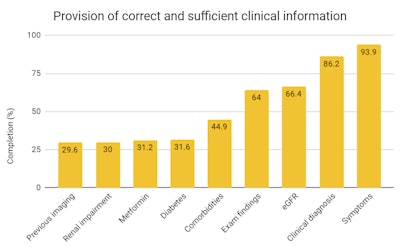 eGFR = estimated glomerular filtration rate.
"Request cards were generally completed poorly," Agarwal told delegates at the 2019 UK Imaging & Oncology Congress in Liverpool. "Symptoms and clinical diagnosis were completed better, [at] 93.9% and 86.2%, respectively. Documentation of previous imaging, metformin use, and renal status were completed poorly at 29.6%, 31.2%, and 30%, respectively."
She also looked for the presence of high-risk features of symptoms, examination, and comorbidities for each patient, and she stratified patients into groups based on low (51%), intermediate (40%), and high risk (9%) of AAS.
Overall, CT scanning was justified in 22 of the 247 patients (8.9%). Only three patients had confirmed aortic pathology on CT scans. The remaining 23 patients with confirmed aortic pathology were classified as low or intermediate risk.
"Justification of scans is dependent on clinical information provided on the request card by the referrer," Agarwal pointed out. "Due to the lack of sufficient documentation, it is difficult to determine true risk of AAS in patients."
She attributed increased CT requests to the following:
- Patients are indicated for CT of the aorta, but requests are inadequately completed due to clinical urgency.
- There is a lack of resources, including equipment and trained staff, to perform ultrasound scans on patients.
- Litigation in healthcare has increased, causing defensive practice in medicine.
- CT aorta scans are useful in excluding AAS and identifying other acute pathology within the thorax and abdomen.
The main limitation of this study was that two request cards were not found on the current research information system, and the authors used the clinical indication listed on the report and included the patients in the sample. Also, one scan was done as part of a trauma protocol, and the findings may be subject to reporting bias, Agarwal and Amonkar conceded.
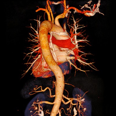
Editor's note: The image used to introduce this article on our homepage consists of a CT angiogram of the aorta at low kV and high mAs with High Power 80 for iodine contrast and potential reduction of required contrast media dosage (tube current = 80 kV/375 mAs, CT dose index volume = 3.45 mGy, dose-length product = 147 mGy-cm). Image courtesy of University Hospital of Erlangen, Germany, and Siemens Healthineers.

