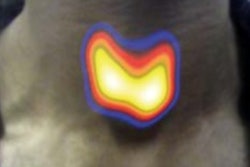
SPECT gamma cameras featuring cadmium zinc telluride-based (CZT) digital detector technology are just what the nuclear cardiologist ordered to diagnose seemingly healthy patients with cardiac symptoms and to help predict their chances for an adverse event, according to a study published online April 27 in the Journal of Nuclear Medicine.
Dutch researchers used the modality to confirm that the degree of abnormal myocardial perfusion imaging (MPI) increased along with the chances of these patients experiencing an adverse event within three years. CZT-based SPECT achieved the results with less radiation exposure for cardiac patients and no degradation in diagnostic image quality.
"Our findings show that MPI acquired with [a] CZT SPECT camera provides excellent prognostic information, with low event rates in patients with normal myocardial perfusion," wrote lead author Dr. Elsemiek Engbers and colleagues at Isala Hospital in Zwolle. "In patients with abnormal SPECT MPI, the extent of abnormality is independently associated with an increased risk of events."
SPECT MPI
Myocardial perfusion imaging with conventional photomultiplier-based SPECT cameras has long been used to diagnose and predict likely outcomes for patients with coronary artery disease. Previous research has shown that when patients have a normal SPECT MPI result, they have an excellent prognosis and an annual adverse cardiac event rate of less than 1%.
More recently, concerns over exposing patients to radiation have prompted the development of more novel gamma cameras with amenities such as CZT solid-state digital detectors and advanced collimator configurations to image the heart more directly.
"Novel cardiac gamma cameras utilizing multipinhole collimation and solid-state detectors offer several advantages compared to the conventional SPECT cameras," the authors wrote. "Imaging time and radiation dose can be reduced, while diagnostic accuracy appears to be unchanged."
There is, however, a paucity of data on the modality's prognostic capabilities, Engbers and colleagues wrote. Therefore, they set out to assess the technology in what they assert is the largest cohort to date (JNM, April 27, 2017).
The researchers enrolled 4,057 consecutive patients (mean age, 61 years ± 11 years) referred between May 2010 and June 2013 who had no history of coronary artery disease (CAD). The patients were referred to the facility for atypical chest pain and dyspnea. Patients with a known history of CAD were excluded from the study.
The subjects initially underwent a stress test with low-dose technetium-99m (Tc-99m) tetrofosmin for SPECT imaging. In cases of an abnormal stress perfusion, additional rest SPECT scans were performed. For pharmacological stress tests, patients received adenosine unless there was a contraindication; as an alternative, they were given dobutamine or regadenoson or rode a stationary bicycle. Rest-imaging patients received a weight-based dose of Tc-99m tetrofosmin.
Clinicians performed both stress and rest SPECT scans 45 to 60 minutes after tracer injection. The time between the stress and rest tests was more than three hours. Scans were conducted on a CZT-based SPECT/CT system (Discovery NM/CT 570c, GE Healthcare), which features 19 stationary CZT detectors that simultaneously acquire 19 cardiac images.
Two experienced nuclear cardiology readers blindly interpreted the images by consensus using a five-point rating system in which "0" was a normal result and "4" indicated the absence of detectable tracer uptake and thus an abnormal result. Perfusion images were reviewed again after both stress and rest SPECT.
The subjects were monitored for primary outcomes of nonfatal myocardial infarction and cardiac mortality, as well as secondary outcomes including late revascularization (more than 90 days after SPECT).
Prognostic prowess
In total, 3,137 subjects (77%) had normal SPECT MPI findings and 920 (23%) had abnormal images.
Of those with abnormal perfusion results, 558 patients (61%) had small total perfusion defects and 362 (39%) had moderate/large total perfusion defects. In addition, 575 patients had ischemic perfusion defects: 353 (61%) had small and 222 (39%) had moderate/large defects.
During the median follow-up of 2.4 years, eight patients were lost, leaving 4,049 patents in the subsequent analysis. The researchers categorized the patients by type of outcome, and those with normal MPI results had low annual event rates of primary (0.2%) and secondary outcomes (0.6%).
By comparison, patients with abnormal SPECT MPI scans and evidence of small, moderate, or large ischemic defects were more likely to have adverse event.
| Annual event rates for patients with abnormal MPI results | ||
| Primary outcome | Secondary outcome | |
| Moderate/large ischemic defect | 1.2% | 4.3% |
| Small ischemic defect | 0.7% | 2.8% |
There were 37 primary events (0.9%) during the follow-up period: 14 cardiac deaths (38%) and 23 nonfatal myocardial infarctions (62%). There were also 116 secondary events (2%), including 79 late revascularizations (68%).
The extent of ischemia was significantly associated with the risk for an event, the researchers found. Having a moderate or large ischemic defect carried the highest risk. Being male, older, or having diabetes were less significant but still risk factors.
| Risk for events after multivariate analysis | ||
| Hazard ratio (95% confidence interval) | ||
| Primary outcome | Secondary outcome | |
| Moderate/large ischemic defect | 4.0 (1.5-10.5) | 12.1 (7.2-20.2) |
| Small ischemic defect | 2.2 (0.9-5.9) | 4.6 (2.8-7.6) |
The next step
"Our study demonstrated that MPI acquired with the CZT SPECT camera has excellent prognostic value in patients suspected for CAD," Engbers and colleagues concluded. "A normal MPI is associated with low event rates and in abnormal MPI, the extent of abnormality is associated with an increased risk of events."
The group suggested that future research be directed toward reducing radiation dose and imaging time.
"This can be achieved by implementing a stress-first imaging protocol, in which rest imaging is omitted when the stress images are normal," they added.


