Major contrast reductions in coronary CT angiography (CCTA) can be achieved across vendor platforms with a couple of simple techniques, according to a Dutch presentation at this month's ECR 2017 in Vienna.
 Dr. Robbert van Hamersvelt from University Medical Center Utrecht.
Researchers from University Medical Center (UMC) Utrecht tested the techniques for cutting contrast use in dual-energy CT (DECT) scanners from three vendors: low kVp in single-source mode, and DECT -- all acquired in a circulation phantom that simulated real physiological conditions. Despite widely varying technologies among the scanners, the results were fairly consistent for each of the two scanning modes.
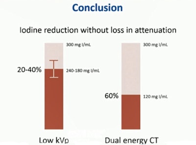
The investigators found that standard CT with the lowest possible kVp settings enables iodine reductions of 20% to 40% without any loss of intravascular attenuation. Using DECT mode, virtual monochromatic imaging (VMI) at 40 keV allows for 60% reductions, or as little as 120 mgI/mL, for all three vendors.
"Iodine reduction without loss of attenuation can be achieved all the way to 20% to 40% on low-kVp scanning -- and down to 60% using dual-energy CT scans," said Dr. Robbert van Hamersvelt, a PhD student in radiology at UMC Utrecht.
Contrast dose matters for some
CCTA is a robust test for assessing the coronary arteries, but its use is limited in patients with impaired renal function due to the risk of contrast-related nephropathy, he said. For these patients, the ideal situation would be to reduce contrast dose while maintaining the same attenuation, a solution that would offer benefits in terms of both safety and costs.
"The objective of our study was to evaluate the feasibility of iodine concentration reduction in DECT from three major vendors, using low kVp and dual-energy CT," van Hamersvelt noted. Models included a dual-source CT (DSCT) scanner (Somatom Force, Siemens Healthineers), a fast kVp switching scanner (Discovery 750HD, GE Healthcare), and a dual-layer detector CT scanner (IQon Spectral, Philips Healthcare).
The team assessed blood flow with a circulation phantom set to mimic a heart rate of 60 bpm, blood pressure 120/80 mmHg, and stroke volume of 60 mL per beat. A series of tubes mimics the coronary arteries. Assessed in the studies were regions of interest (ROIs) of the left main coronary artery, ROIs of the ascending and descending aorta.
They used six different iodine concentrations starting with 300 mL of iodine -- standard in the authors' institution -- and reduced it with 20% increments up to a 90% reduction (300, 240, 180, 60, 30 mGI/mL) using a fixed injection protocol (flow rate 6 mL/s, volume 40 mL, and a fixed injection time of 6.7 seconds).
Image acquisition began in single-source mode using the lowest kVp possible for each vendor for each of the three models (DSCT, 70 kVp; fast kV switching scanner, 80 kVp; dual-layer scanner, 80 kVp). This was followed by scanning using the DECT settings, van Hamersvelt said.
Subsequently images were acquired in DECT mode (DSCT 80 to 150 Sn; fast kV switching 80 to 140 Sn; dual-layer 140 kVp). VMI ranging from 40 keV to 120 keV were reconstructed for each DECT acquisition. CT attenuation values (HU) were obtained from regions of interest in the coronary artery and aorta.
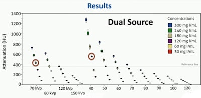
The results show that without reducing attenuation (results above the reference line in the three graphs above), iodine concentration can be reduced by 20% to 40% with low kVp scanning, and by 60% for dual-energy CT scanning, van Hamersvelt concluded.
 DSCT iodine reduction without loss.
Study limitations include the use of a phantom, and the lack of diagnostic evaluation of the scanning results or image quality, he said.


